
Atlas of ultrasound images - scanner Accuvix-XQ
In this section of ultrasound image galery the sonograms received by scanner Accuvix XQ (Medison) are represented.
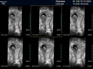
Bicornate uterus, MSV.
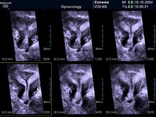
Bicornate uterus, MSV.
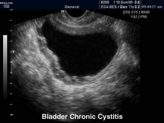
Bladder - chronic cystitis, B-mode.
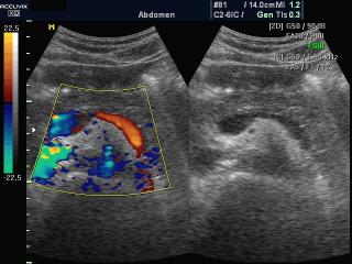
Blood flow in epigastrio, color doppler (videо).
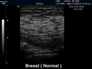
Breast - norm, B-mode.
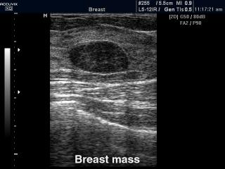
Breast nodule, B-mode.
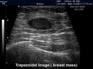
Breast nodule, trapezoidal mode.
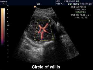
Circle of Willis, power doppler.
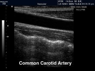
Common carotid artery, B-mode.
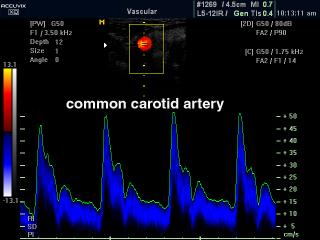
Common carotid artery, CFM & PW.
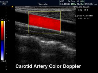
Common carotid artery, color doppler.
Epignatus - defect of fetal`s development, 3D.
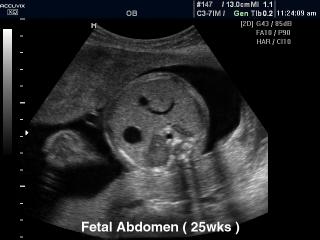
Fetal abdomen - 25 weeks, B-mode.
Fetal brain - expansion of the lateral ventricles, MR.
Fetal diaphragm, B-mode.
Fetal face and umbilical cord, 3D (videо).
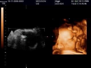
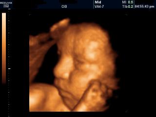
Fetal face, 3D (videо).
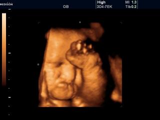
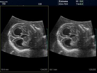
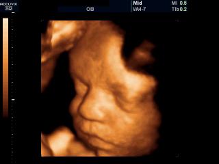
Fetal face, 3D.
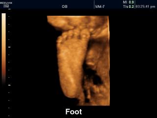
Fetal foot, 3D.
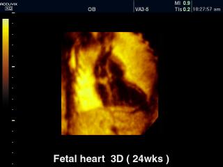
Fetal heart - 24 weeks, 3D.
Fetal heart - aortic arch, color doppler (videо).
Fetal heart - LVOT, color doppler (videо).
Fetal heart - RVOT, color doppler (videо).
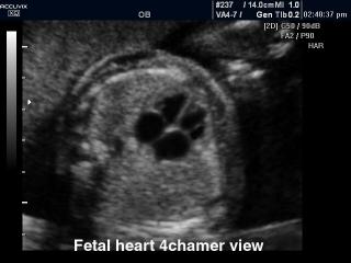
Fetal heart (4 chamber view), B-mode.
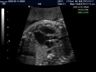
Fetal heart (5 chamber view), B-mode.
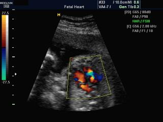
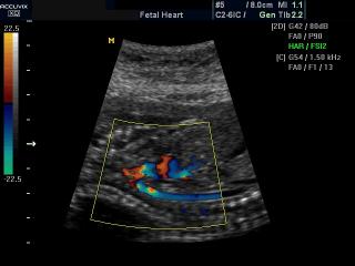
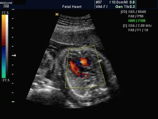
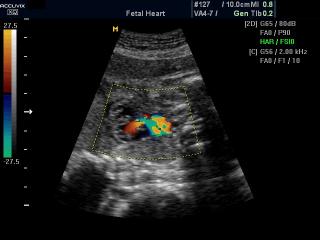
Fetal heart, color doppler (videо).
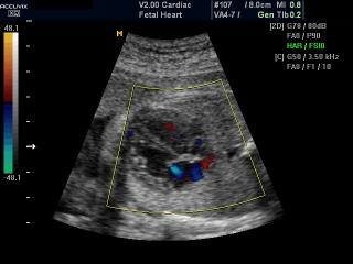
Fetal heart, color doppler (videо).
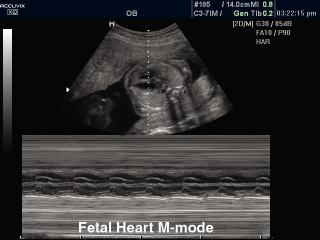
Fetal heart, M-mode.
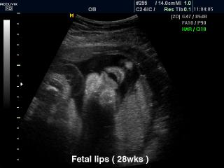
Fetal lips - 28 weeks, B-mode.
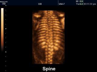
Fetal spine, 3D.

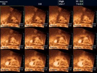
Fetal stomach, VOCAL.
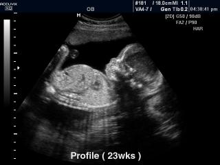
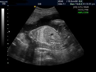
Fetus - 23 weeks, B-mode.
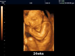
Fetus - 24 weeks, 3D.
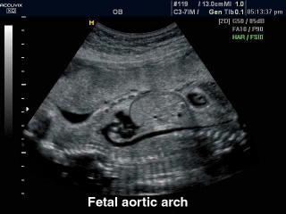
Fetus - arotic arch, B-mode.
Fetus - cleft lip, MSV.
Fetus - nuchal translucency, MSV.
The information about equipment: scanner Accuvix XQ (full description in Russian).
The description of ultrasound examinations in russian (эхография) and in english are situated in the database. The description of the diagnostic in the english language is closer to latin and doctors are likely to prefer this variant.