
Атлас УЗИ - акушерство
В разделе "Акушерство" атласа представлены результаты ультразвуковых исследований беременных на разных сроках. Здесь вы можете посмотреть изображения внутренних органов, мозга, сердца и пола плода, эхограммы многоплодной беременности, кровотока в плаценте и пуповине, пороков развития и др.
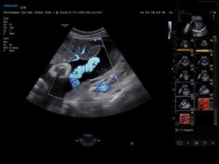
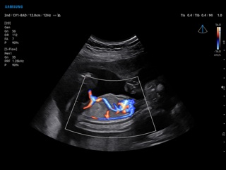
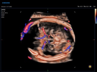
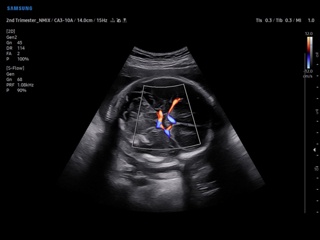
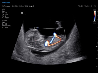
V6. Пуповина плода, MV-Flow.
V6. Пуповина плода, CrystalVue Flow.
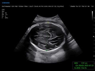
V6. Головной мозг плода, BiometryAssist.
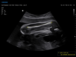
V6. Бедро плода, ViewAssist.
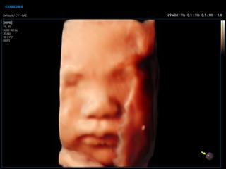
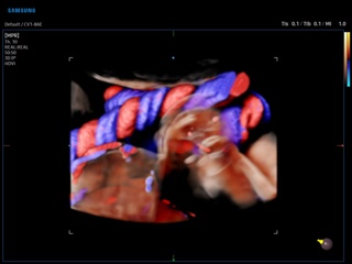
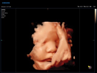
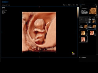
V6. Лицо плода, RealisticVue, 3D.
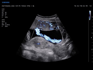
V7. Плод, LumiFlow.
V7. Головной мозг плода, MV-Flow.
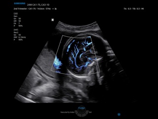
V7. Позвоночник плода, CrystalVue, 3D.
V7. Головной мозг плода, CrystalVue Flow, 3D.
V7. Лицо плода, RealisticVue, 3D.
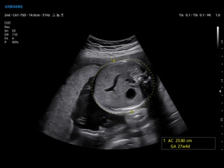
V7. Плод, BiometryAssist.
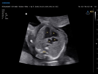
V7. Сердце плода (4-камерная позиция), ViewAssist.
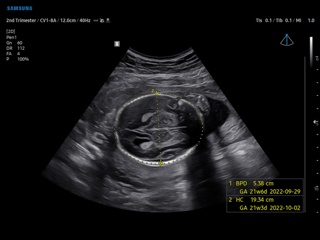
V8. Голова плода, BiometryAssist.
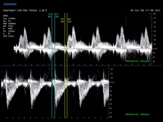
V8. Сердце плода, RV MPI.
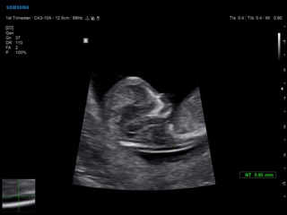
V8. Плод - измерение ТВП, BiometryAssist.
V8. Сердце плода, S-Flow и LumiFlow.
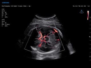
V8. СМА плода, S-Flow.
V8. Плод - ранний срок беременности, Realistic Vue, 3D.
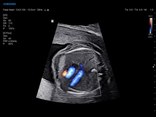
V8. Пуповина плода, MV-Flow.
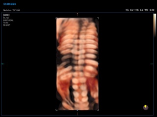
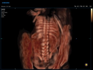
V8. Скелет плода, CrystalVue, 3D.
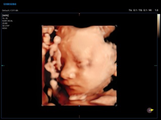
V8. Лицо плода, RealisticVue, 3D.
V8. Плод, S-Flow и LumiFlow.
V8. Головной мозг плода, MV-Flow.
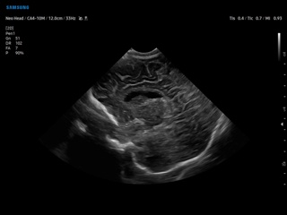
V8. Голова новорожденного.
Страницы: 01 02 03 04 05 06 следующая
В разделе "Эхография в акушерстве" журнала "SonoAce-Ultrasound" Вы можете ознакомиться с публикациями врачей по теме.
В базе данных имеется описание ультразвуковых исследований на русском и английском языках (sonography). Описание диагноза на английском языке ближе к латыни и, вероятно, врачи предпочтут именно этот вариант.