
Atlas of ultrasound images (pg. 29)
The atlas of echograms is created for demonstrating the capabilities of Samsung Medison ultrasound scanners. The main part of ultrasound images is received from Korea, new echograms - from users of Samsung Medison scanners in Russia: institutes, diagnostic medical centres and private practicing doctors. The material is recommended for specialists of ultrasound diagnostics.
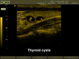
SonoAce-Pico. Thyroid cyst, B-mode.
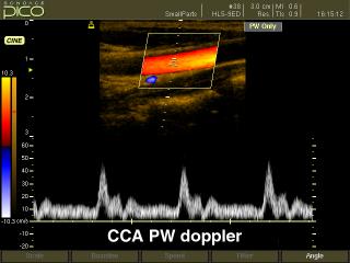
SonoAce-Pico. Common carotid artery, CFM & PW.
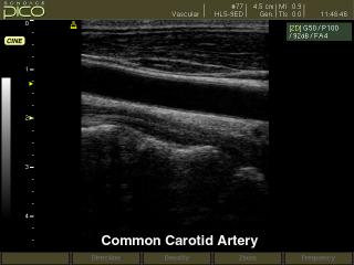
SonoAce-Pico. Common carotid artery, B-mode.
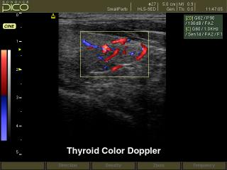
SonoAce-Pico. Thyroid, color doppler.
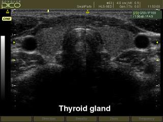
SonoAce-Pico. Thyroid, B-mode.
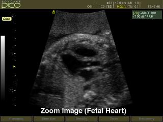
SonoAce-Pico. Fetal heart, B-mode.
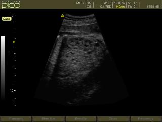
SonoAce-Pico. Fetal kidney, B-mode.
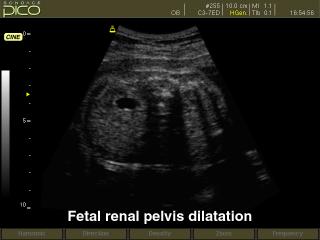
SonoAce-Pico. Fetal renal pelvis dilatation, B-mode.
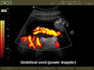
SonoAce-Pico. Umbilical cord, power doppler.
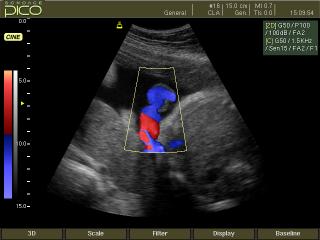
SonoAce-Pico. Umbilical cord insertion, color doppler.
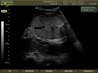
SonoAce-Pico. Fetal abdomen, B-mode.
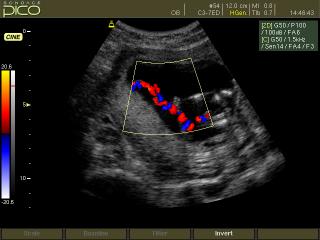
SonoAce-Pico. Umbilical cord, color doppler.
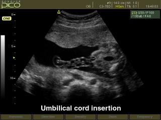
SonoAce-Pico. Umbilical cord insertion, B-mode.
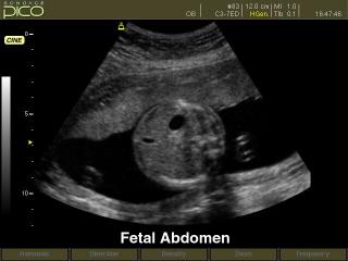
SonoAce-Pico. Fetal abdomen, B-mode.

SonoAce-Pico. Circle of Willis, power doppler.
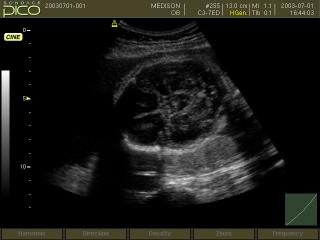
SonoAce-Pico. Fetal head, B-mode.
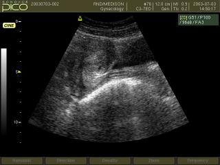
SonoAce-Pico. Uterus, B-mode.
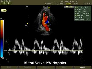
SonoAce-Pico. Mitral valve, CFM & PW.
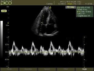
SonoAce-Pico. Mitral valve, PW.
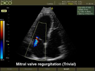
SonoAce-Pico. Mitral valve regurgitation, color doppler.
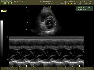
SonoAce-Pico. Mitral valve, M-mode.
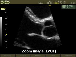
SonoAce-Pico. Left ventrical - outflow track, B-mode.
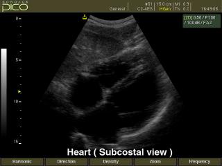
SonoAce-Pico. Heart (subcostal view), B-mode.
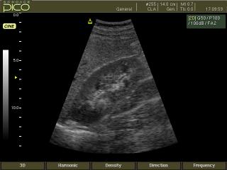
SonoAce-Pico. Kidney, B-mode.
The description of ultrasound examinations in russian (эхография) and in english are situated in the database. The description of the diagnostic in the english language is closer to latin and doctors are likely to prefer this variant.