
Atlas of ultrasound images - obsterics (pg. 12)
In the section "Оbsterics" of atlas the results of ultrasonic examinations of pregnant women with different durations of gestation are represented. Here you can see images of internally organs, cerebrum, cordis and the sex of the fetus, the sonograms of multiple pregnancy, the blood flow in placenta and umbilical cord, defects of fetal`s development, etc.
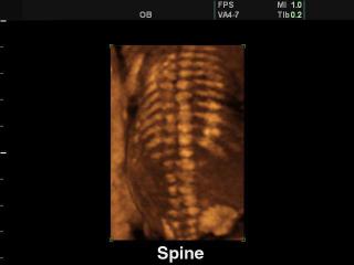
SonoAce-8000. Fetal spine, 3D.
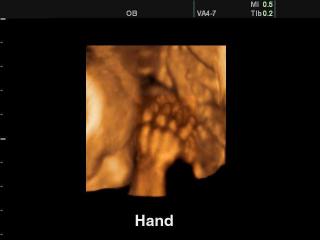
SonoAce-8000. Fetal hand, 3D.
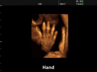
SonoAce-8000. Fetal hand, 3D.
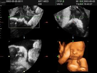
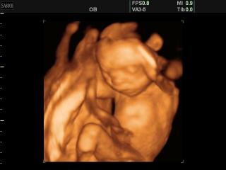
SonoAce-8000. Fetus, 3D.
SonoAce-8000. Fetus - 20 weeks, 3D.
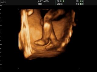
SonoAce-8000. Fetal foots, 3D.
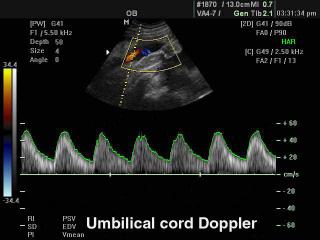
SonoAce-8000. Umbilical cord, CFM & PW.
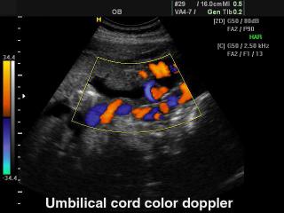
SonoAce-8000. Umbilical cord, сolor doppler.
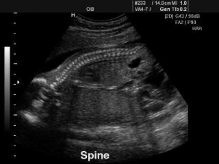
SonoAce-8000. Fetal spine, B-mode.
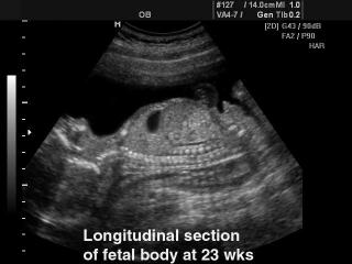
SonoAce-8000. Fetus - 23 week, B-mode.
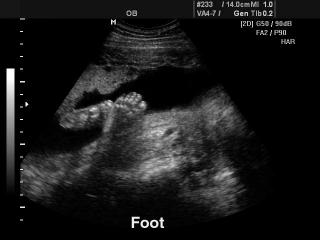
SonoAce-8000. Fetal foot, B-mode.
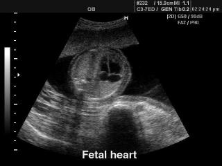
SonoAce-8000. Fetal heart, B-mode.
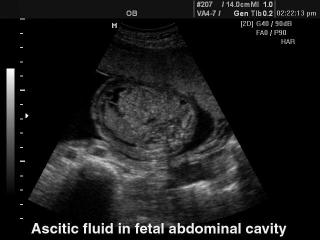
SonoAce-8000. Fetal abdomen ascites, B-mode..
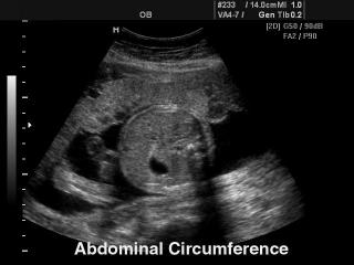
SonoAce-8000. Fetus - abdominal circumference, B-mode.
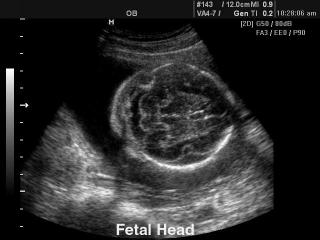
SonoAce-8000. Fetal brain, B-mode.
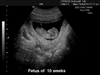
SonoAce-8000. Fetus - 10 weeks, B-mode.
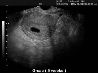
SonoAce-8000. Gestational sac - 5 weeks, B-mode.
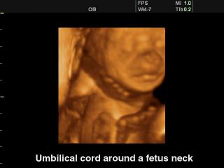
SonoAce-8000. Umbilitical cord around a fetus neck, 3D.
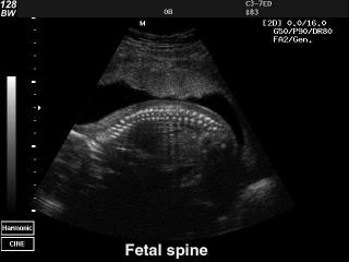
SonoAce-128BW. Fetal spine, B-mode.
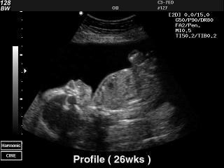
SonoAce-128BW. Fetus - 26 weeks, B-mode.
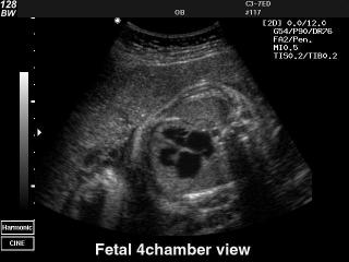
SonoAce-128BW. Fetal heart (4 chamber view), B-mode.
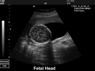
SonoAce-128BW. Fetal head, B-mode.
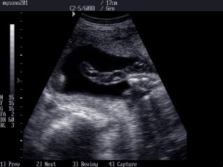
MySono-201. Umbilical cord, B-mode.
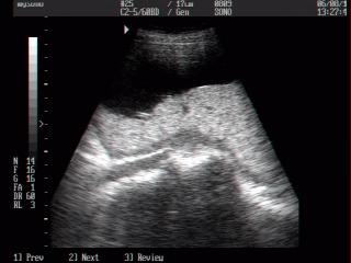
MySono-201. Placenta, B-mode.
The description of ultrasound examinations in russian (эхография) and in english are situated in the database. The description of the diagnostic in the english language is closer to latin and doctors are likely to prefer this variant.