
Atlas of ultrasound images (pg. 19)
The atlas of echograms is created for demonstrating the capabilities of Samsung Medison ultrasound scanners. The main part of ultrasound images is received from Korea, new echograms - from users of Samsung Medison scanners in Russia: institutes, diagnostic medical centres and private practicing doctors. The material is recommended for specialists of ultrasound diagnostics.
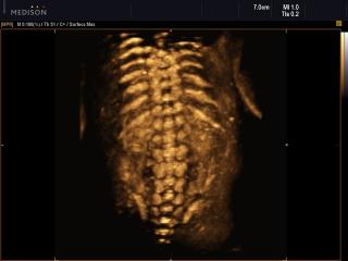
Accuvix-XG. Fetal spine, HDVI, 3D.
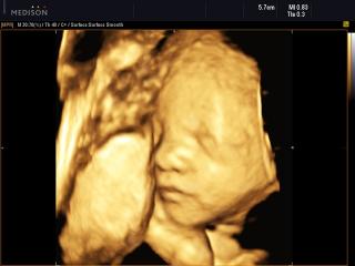
Accuvix-XG. Fetal face, 3D.
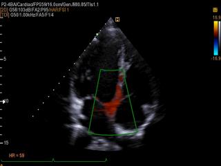
EKO7. Interventricular septum, TDI color doppler.
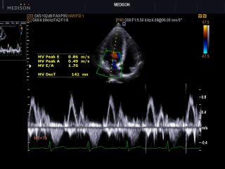
EKO7. Transmitral flow, CFM & PW.
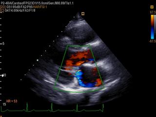
EKO7. Mitral regurgitation, color doppler.
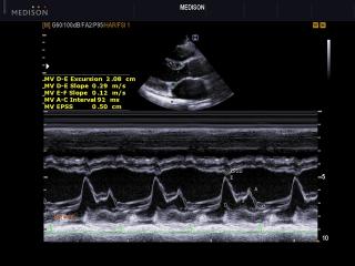
EKO7. Mitral valve, M-mode.
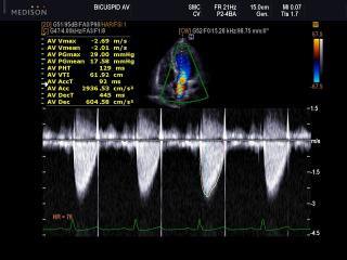
EKO7. Transaortic flow, CFM & CW.
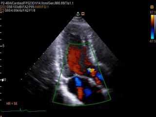
EKO7. Mitral regurgitation and aortic regurgitation, color doppler.
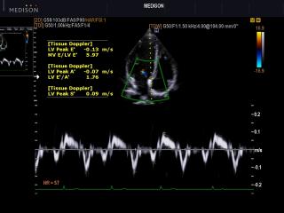
EKO7. Interventricular septum, TDI spectral doppler.
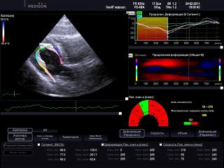
EKO7. Left ventricular longitudinal, 2D Strain and endocardial vectors.

EKO7. Left ventricular longitudinal, 2D Strain and acoustic markers.
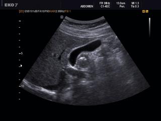
EKO7. Gall bladder, B-mode.
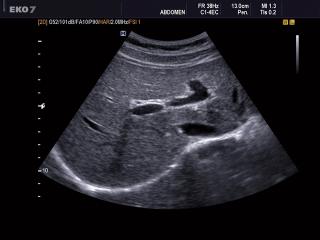
EKO7. Liver, B-mode.
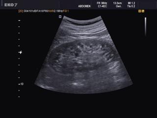
EKO7. Kidney, B-mode.
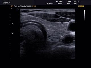
EKO7. Thyroid, B-mode.
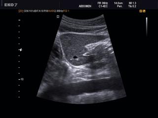
EKO7. Liver - left lobe, B-mode.
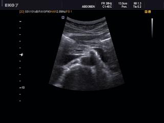
EKO7. Pancreas, B-mode.
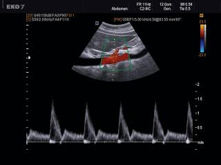
EKO7. Celiac artery, CFM & PW.
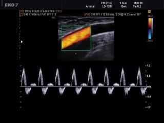
EKO7. Common femoral artery, CFM & PW.
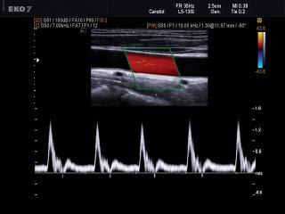
EKO7. Internal carotid artery, CFM & PW.
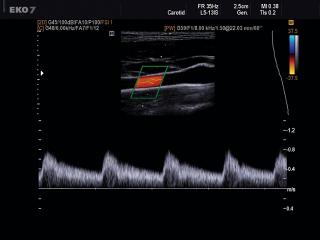
EKO7. Common carotid artery, CFM & PW.
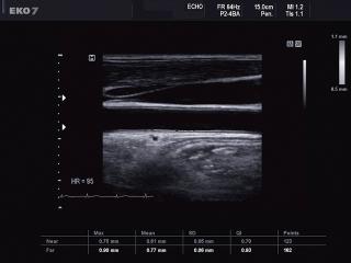
EKO7. Common carotid artery, Auto IMT.
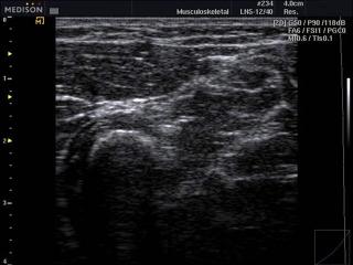
SonoAce-R3. Soft tissue, B-mode.
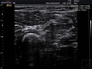
SonoAce-R3. Soft tissue, Quick-FSI.
The description of ultrasound examinations in russian (эхография) and in english are situated in the database. The description of the diagnostic in the english language is closer to latin and doctors are likely to prefer this variant.