
Atlas of ultrasound images (pg. 33)
The atlas of echograms is created for demonstrating the capabilities of Samsung Medison ultrasound scanners. The main part of ultrasound images is received from Korea, new echograms - from users of Samsung Medison scanners in Russia: institutes, diagnostic medical centres and private practicing doctors. The material is recommended for specialists of ultrasound diagnostics.
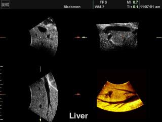
SonoAce-9900. Liver, 3D reconstruction.
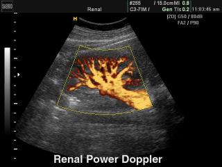
SonoAce-9900. Kidney, power doppler.
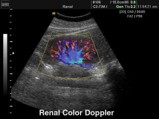
SonoAce-9900. Kidney, color doppler.
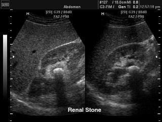
SonoAce-9900. Renal stone, B-mode.
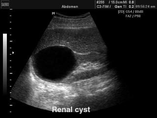
SonoAce-9900. Renal cyst, B-mode.
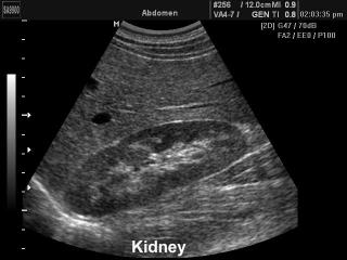
SonoAce-9900. Kidney, B-mode.
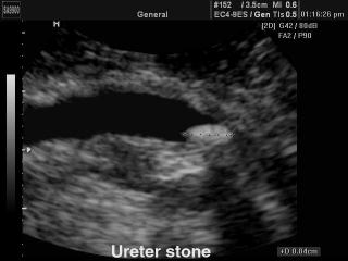
SonoAce-9900. Ureter stone, B-mode.

SonoAce-9900. Spleen - splenomegaly, B-mode.
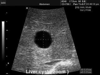
SonoAce-9900. Liver cyst, B-mode.
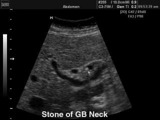
SonoAce-9900. Stone in the neck of gallbladde, B-mode.
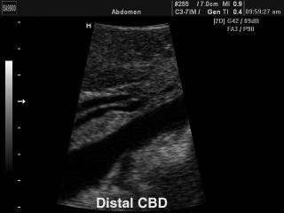
SonoAce-9900. Liver - distal common bile duct, B-mode.
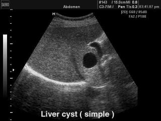
SonoAce-9900. Liver - simple cyst, B-mode.
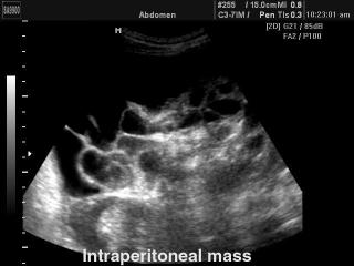
SonoAce-9900. Intestines - intraperitoneal induration, B-mode..
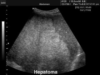
SonoAce-9900. Liver - hepatoma, B-mode.
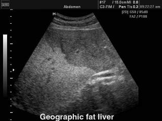
SonoAce-9900. Liver steatosis, B-mode.
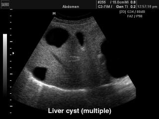
SonoAce-9900. Liver polycystic, B-mode.
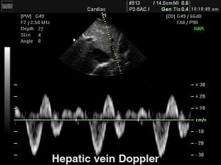
SonoAce-9900. Hepatic vein, PW.
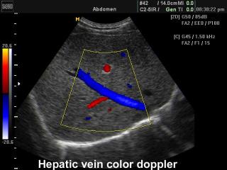
SonoAce-9900. Hepatic vein, color doppler.
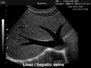
SonoAce-9900. Liver and hepatic veins, B-mode.
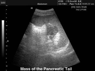
SonoAce-9900. Pancreatic tail mass, B-mode.
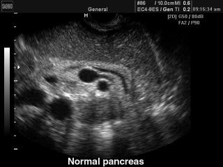
SonoAce-9900. Pancreas - norm, B-mode.
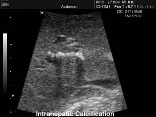
SonoAce-9900. Intrahepatic calcification, B-mode.
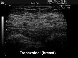
SonoAce-8000. Breast, trapezoidal B-mode.
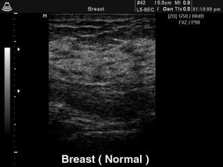
SonoAce-8000. Breast - norm, B-mode.
The description of ultrasound examinations in russian (эхография) and in english are situated in the database. The description of the diagnostic in the english language is closer to latin and doctors are likely to prefer this variant.