
Atlas of ultrasound images (pg. 34)
The atlas of echograms is created for demonstrating the capabilities of Samsung Medison ultrasound scanners. The main part of ultrasound images is received from Korea, new echograms - from users of Samsung Medison scanners in Russia: institutes, diagnostic medical centres and private practicing doctors. The material is recommended for specialists of ultrasound diagnostics.

SonoAce-8000. Common carotid artery, CFM & PW.
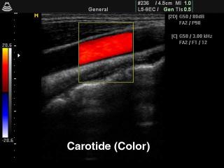
SonoAce-8000. Common carotid artery, color doppler.
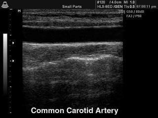
SonoAce-8000. Common carotid artery, B-mode.
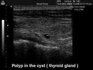
SonoAce-8000. Thyroid polyp in the cyst, B-mode.
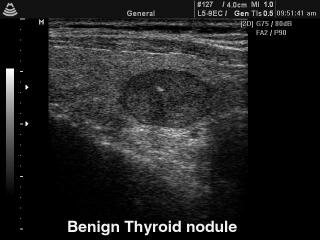
SonoAce-8000. Thyroid benign nodule, B-mode.
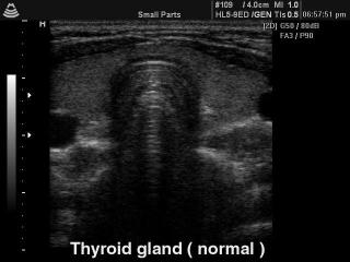
SonoAce-8000. Thyroid - norm, B-mode.
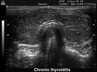
SonoAce-8000. Chronic thyroiditis, B-mode.
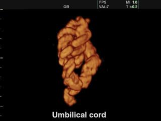
SonoAce-8000. Umbilical cord, 3D.
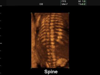
SonoAce-8000. Fetal spine, 3D.
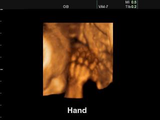
SonoAce-8000. Fetal hand, 3D.
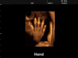
SonoAce-8000. Fetal hand, 3D.
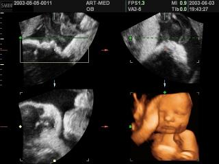
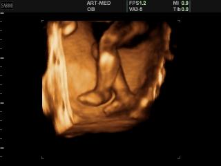
SonoAce-8000. Fetus, 3D.
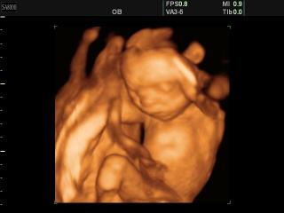
SonoAce-8000. Fetus - 20 weeks, 3D.
SonoAce-8000. Fetal foots, 3D.
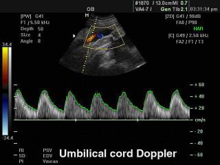
SonoAce-8000. Umbilical cord, CFM & PW.
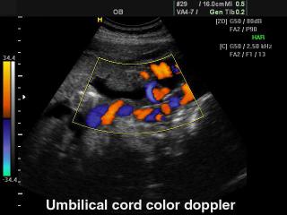
SonoAce-8000. Umbilical cord, сolor doppler.
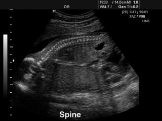
SonoAce-8000. Fetal spine, B-mode.
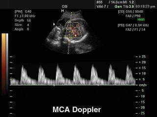
SonoAce-8000. Fetal middle cerebral artery, PD & PW.
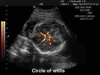
SonoAce-8000. Circle of Willis, power doppler.
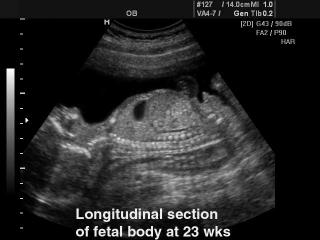
SonoAce-8000. Fetus - 23 week, B-mode.
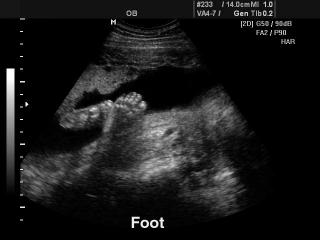
SonoAce-8000. Fetal foot, B-mode.
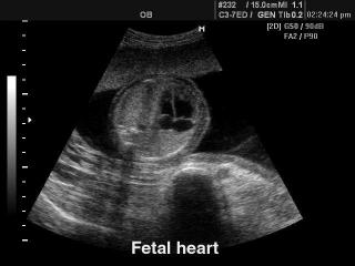
SonoAce-8000. Fetal heart, B-mode.
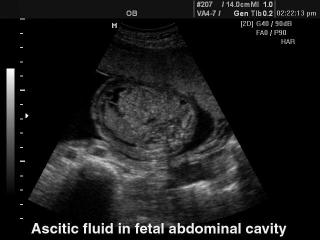
SonoAce-8000. Fetal abdomen ascites, B-mode..
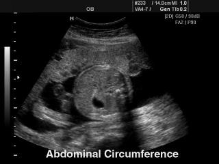
SonoAce-8000. Fetus - abdominal circumference, B-mode.
The description of ultrasound examinations in russian (эхография) and in english are situated in the database. The description of the diagnostic in the english language is closer to latin and doctors are likely to prefer this variant.