
Atlas of ultrasound images
The atlas of echograms is created for demonstrating the capabilities of Samsung Medison ultrasound scanners. The main part of ultrasound images is received from Korea, new echograms - from users of Samsung Medison scanners in Russia: institutes, diagnostic medical centres and private practicing doctors. The material is recommended for specialists of ultrasound diagnostics.
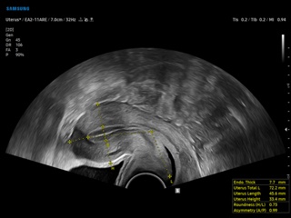
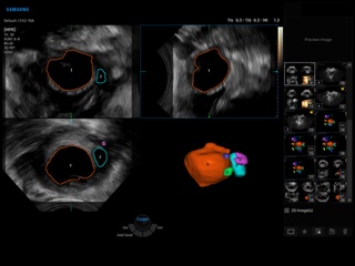
V6. Uterus, UterineAssist.
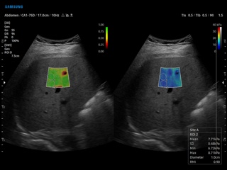
V6. Liver, S-Shearwave.
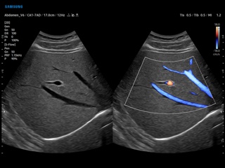
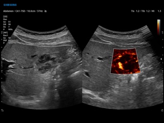
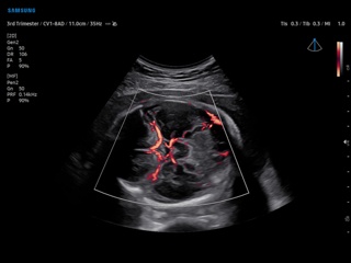
V6. Renal artery, S-Flow & LumiFlow.
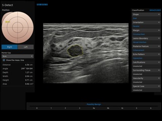
V6. Breast, S-Detect.
V6. Umbilical cord, MV-Flow.
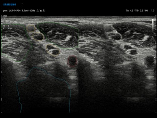
V6. Brachial plexus, NerveTrack.
V6. Liver, EzHRI.
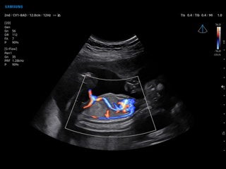
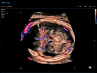
V6. Umbilical cord, CrystalVue Flow.
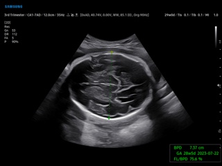
V6. Fetal brain, BiometryAssist.
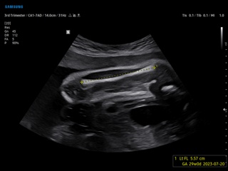
V6. Fetal femur, ViewAssist.
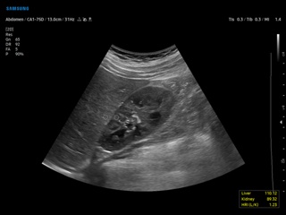
V6. Hepatic vein, S-Flow.
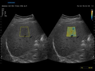
V6. Hepatic steatosis, QUS (TAI+TSI).
V6. Ovarian follicles, 5D Follicle.
V6. Liver hemangioma, MV-Flow.
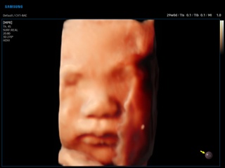
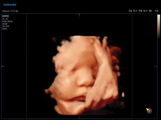
V6. Fetal face, RealisticVue, 3D.
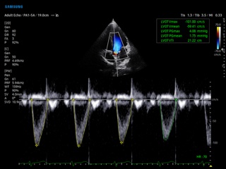
V6. Left ventricular outflow tract, HeartAssist.
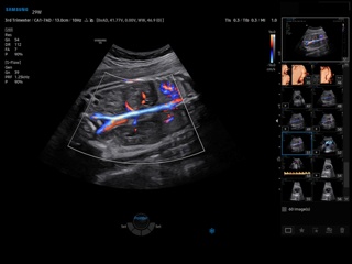
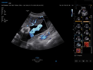
V7. Fetus, LumiFlow.
V7. Fetal brain, MV-Flow.
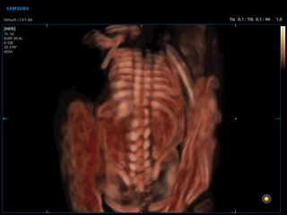
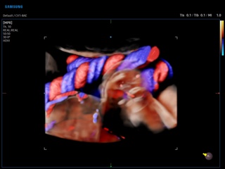
V7. Fetal spine, CrystalVue, 3D.
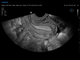
V7. Uterus, B-mode.
V7. Fetal brain, CrystalVue Flow, 3D.
V7. Fetal face, RealisticVue, 3D.
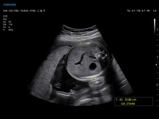
V7. Fetus, BiometryAssist.
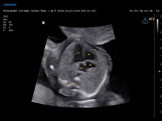
V7. Fetal heart (4 chamber view), ViewAssist.
The description of ultrasound examinations in russian (эхография) and in english are situated in the database. The description of the diagnostic in the english language is closer to latin and doctors are likely to prefer this variant.