
Atlas of ultrasound images - gynecology (pg. 2)
In the section "Gynecology" of atlas the results of ultrasound examinations of female maladies are represented. Here you can see images of uterus and ovaries, fallopian tubes and urinary bladder, the sonograms of ectopic pregnancy, endometrial polyp, myoma, cystic lesion, bicornuate uterus, etc.
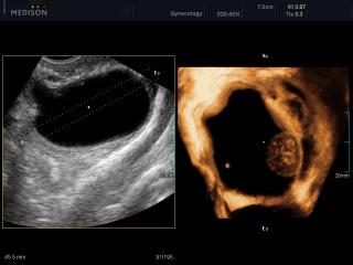
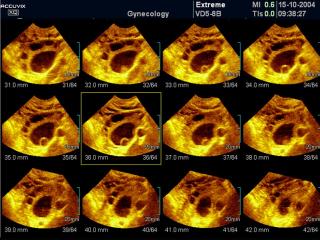
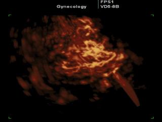
Accuvix-V20. Ovarian mass, OVIX, 3D.
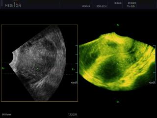
Accuvix-V20. Uterus, OVIX & 3D.
Accuvix-XQ. Uterus - oblique view, B-mode.
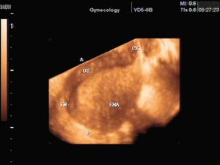
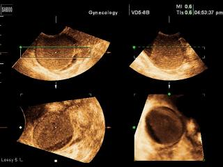
Accuvix-XQ. Uterus, OVIX & 3D.
Accuvix-XQ. Ovary - cyst, MSV.
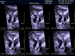
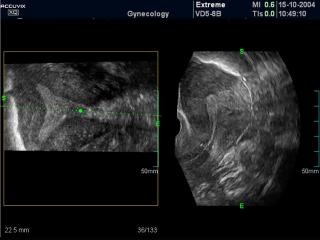
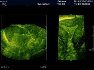
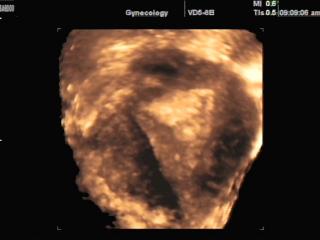
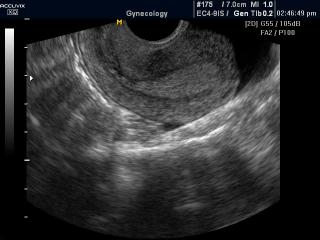
Accuvix-XQ. Bicornate uterus, MSV.
Accuvix-XQ. Bicornate uterus, MSV.
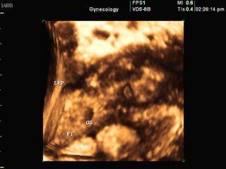
SonoAce-8000. Uterus - subchorionic bleeding, B-mode.
SonoAce-8000. Ectopic pregnancy - fallopian tube dilatation, 3D.
SonoAce-8000. Uterus - subchorionic bleeding, 3D.
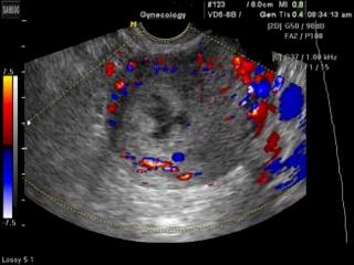
SonoAce-8000. Uterus - subchorionic bleeding, color doppler.
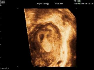
SonoAce-8000. Ectopic pregnancy, 3D.
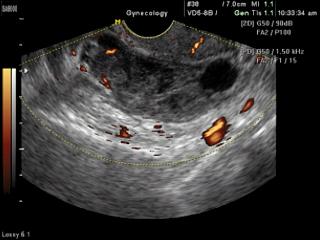
SonoAce-8000. Ectopic pregnancy, power doppler.
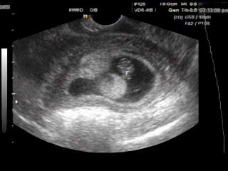
SonoAce-8000. Ectopic pregnancy, B-mode.
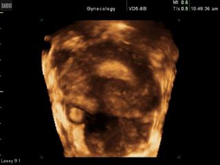
SonoAce-8000. Myometrium, 3D.

SonoAce-8000. Uterine myoma, color doppler, 3D.
SonoAce-8000. Uterine myoma, power doppler, 3D.
SonoAce-8000. Endometrial polyp, 3D.
SonoAce-8000. Ovarian cyst, 3D.
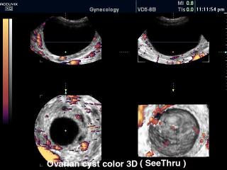
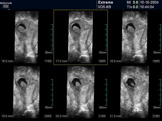
Accuvix-XQ. Ovarian cyst, power doppler & 3D.
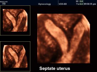
Accuvix-XQ. Septate uterus, 3D.
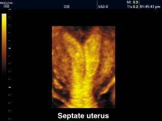
Accuvix-XQ. Septate uterus, 3D.
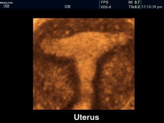
Accuvix-XQ. Uterus, 3D.
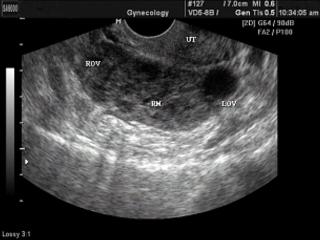
Accuvix-XQ. Uterus, B-mode.
The description of ultrasound examinations in russian (эхография) and in english are situated in the database. The description of the diagnostic in the english language is closer to latin and doctors are likely to prefer this variant.