УЗИ аппарат SonoAce-Pico (Medison, снят с производства)
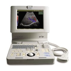
SonoAce Pico - портативный ультразвуковой аппарат компании Medison с цветным допплером и кардиопакетом для исследования сердца плода.
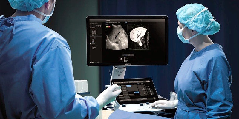
Это современный аппарат для цифрового цветного ультразвукового сканирования. Обладая практически всеми диагностическими возможностями традиционных стационарных сканеров, он является портативным. Благодаря полностью цифровой технологии формирования луча и обработки сигнала, сканер SonoAce Pico позволяет получать изображения с наилучшим для приборов этого класса разрешением. В дополнение к таким технологиям, как формирование трехмерного изображения в ручном режиме и применение широкополосных мультичастотных датчиков, SonoAce Pico имеет функцию формирования трапецеидального изображения, обеспечивающую увеличение поля зрения при исследовании малых органов, возможность использования микроконвексного датчика, а также комплект программ для кардиологических исследований.
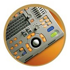
Базовая комплектация: сканер SonoAce Pico (монитор 10,4"; встроенные модули: 2-я гармоника, FreeHand 3D, кардиопакет для исследования сердца плода, SonoView Lite; встроенная клавиатура с трекболом), флакон геля 250 мл и руководство оператора.
Опции для аппарата SonoAce Pico: устройства хранения информации (USB флеш-карта или магнитооптика), система DICOM.
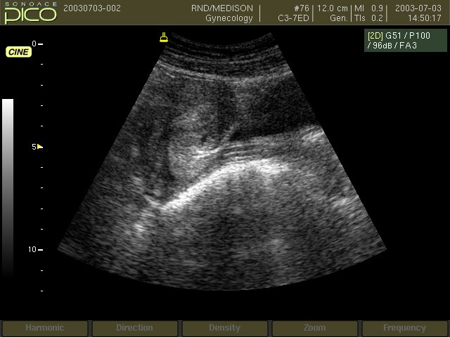
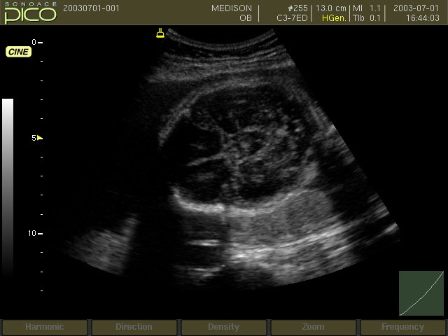
Области применения: акушерство и гинекология, абдоминальные исследования и маммология, урология и эхокардиография, поверхностно расположенные органы и исследования сосудов, мускуло-скелетные исследования, а также педиатрия.
Основные характеристики УЗ аппарата SonoAce-Pico
- Портативный ультразвуковой сканер (357x320x204 мм в собранном виде, вес около 10 кг).
- LCD монитор - 10,4".
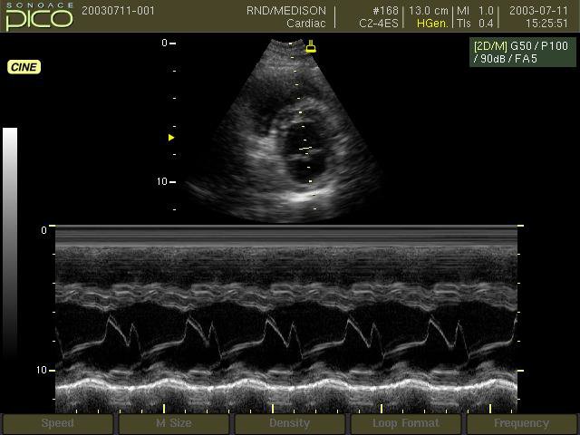
- Режимы сканирования: B, 2B, 4B, M, B+M;
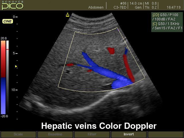
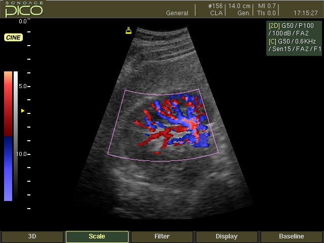
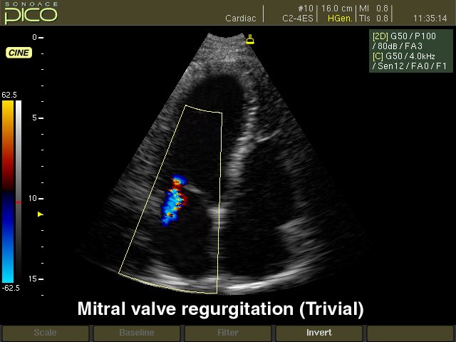
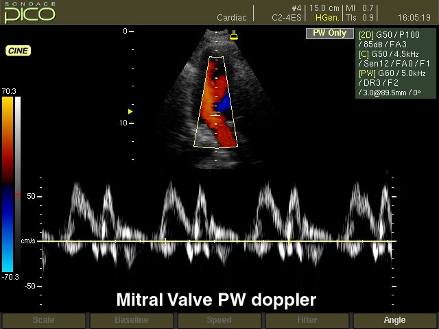
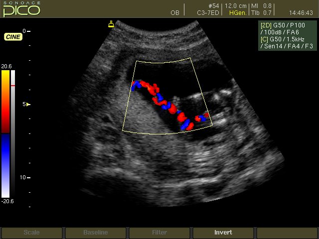
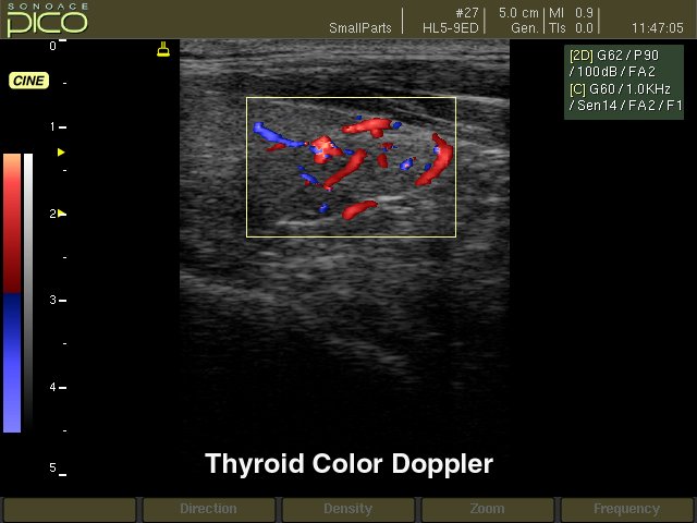
- CFM - цветное допплеровское картирование;
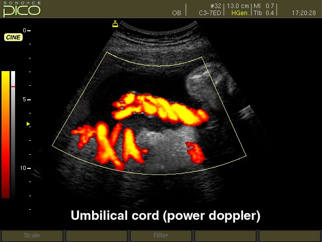
- PD - энергетический допплер;
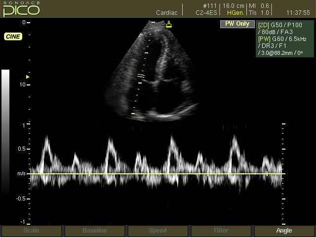
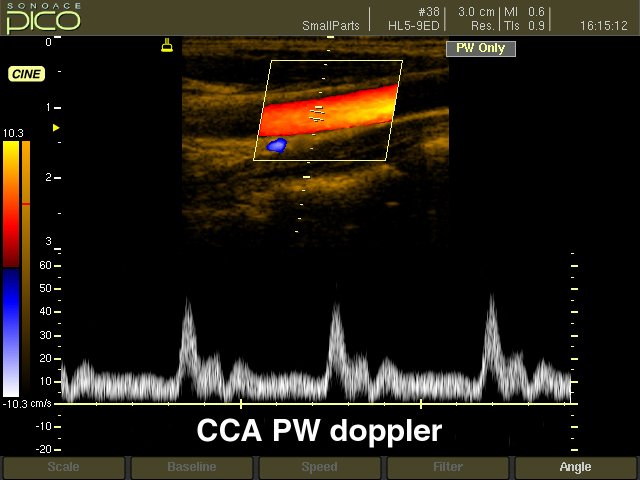
- PW - импульсный допплер;
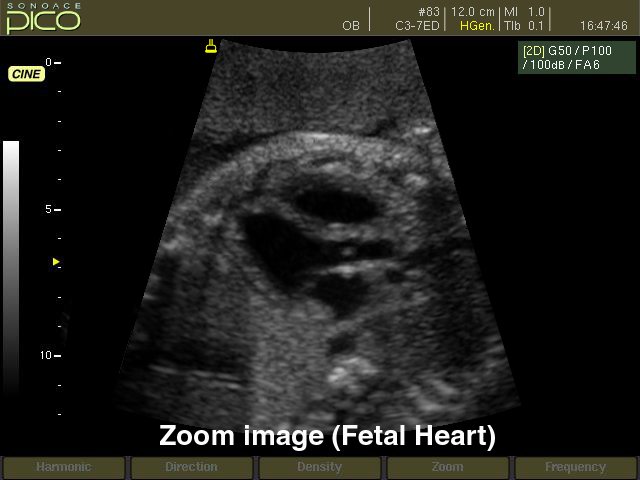
- дуплексный и триплексный режим; трапецевидный режим сканирования для линейных датчиков. - Кардиопакет для исследования сердца плода (входит в базовую комплектацию).
- ЭКГ модуль (входит в базовую комплектацию).
- Глубина сканирования до 30 см.
- Разьемы для одновременного подключения 2-х датчиков.
- Увеличение в реальном масштабе времени, кинопамять, эргономичная клавиатура, мультичастотные датчики.
- Система SonoView Lite - система архивации и дальнейшего просмотра статических эхограмм и динамических клипов.
- Система DICOM (опция) - возможность сетевой интеграции с PACS-системами поддерживающими стандарт DICOM (например, для архивации или печати ультразвуковых эхограмм на оборудовании других производителей медтехники).
Инновационные технологии
- Multi-beam (мульти-луч) - технология цифрового формирования луча (устранение многократного отражения, нелинейного ослабления и неточного времени задержки в отличие от аналоговых систем).
- OTI (Optimum Tissue Imaging) - технология получения оптимального изображения тканей, благодаря коррекции скорости. Функция при помощи которой пользователь может выбрать оптимальную скорость для каждой области исследований, тем самым получая одновременно высокое качество изображений различных видов тканей, таких как жир, мышцы или паренхима печени.
- THI (Tissue Harmonic Imaging), тканевая или 2-я гармоника - повышает качество изображения линейное и контрастное разрешение у трудно визуализируемых пациентов. Данная технология предполагает использование широкополосных датчиков и приемного тракта повышенной чувствительности. Дает преимущество при исследовании пациентов с повышенным весом.
- OHI (Optimized Harmonic Imaging) - объединяет две предыдущие технологии и предназначена для особо трудных для визуализации случаев.
- FINE (Filtered Image for Noise reduction & Edge enhancement) - программа фильтрации ультразвукового изображения. Обеспечивает лучшую контрастность контуров и уменьшает уровень шумов.
- CAFE (Compound Automatic Flash Elemination) - обеспечивает зависимую от используемого режима нелинейную фильтрацию для удаления цветных точек на изображении, возникающих из за мерцающих артефактов. Создает улучшенную визуализацию кровотока во всех допплеровских режимах.
Пакеты ультразвуковых диагностических программ
- Расчет возраста плода с помощью различных измерений: бипариетального диаметра, длины бедренной кости, копчико-теменного размера, диаметра околоплодного мешка, окружности головы, окружности живота и диаметра живота - возможность оценки возраста плода от 4 недель до родов. Расчет веса плода по методам Shepard, Hadlock, Merz, Osaka University. Расчет предпологаемой даты родов по последней менструации. Автоматическое построение графиков роста и веса плода. Просмотр и изменение таблиц возраста плода.
- Кардиологические измерительные программы: оценка митрального и аортального клапанов; оценка левого желудочка по формулам CUBED, POMBO, TEICHHOLZ и в B-режиме: ESP, EBP, BUL, MSR.
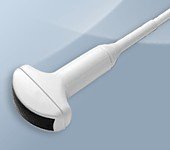
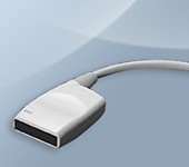
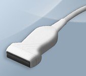
Датчики для УЗ аппарата SonoAce-Pico
Конвексные датчики
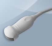
Конвексный датчик 2-4 МГц (микроконвексный)
Кардиология, абдоминальные исследования (печень, желчный пузырь, поджелудочная железа, селезенка, глубокие сосуды), почки.
Конвексный датчик 2-5 МГц
Акушерские исследования (плод, сердце плода), гинекология (матка, яичники), абдоминальные исследования (печень, желчный пузырь, поджелудочная железа, селезенка, глубокие сосуды), почки.
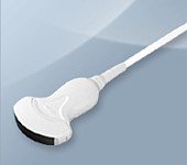
Конвексный датчик 3-7 МГц
Акушерские исследования (плод, сердце плода), гинекология (матка, яичники), абдоминальные исследования (печень, желчный пузырь, поджелудочная железа, селезенка, глубокие сосуды), почки.
Конвексный датчик 4-9 МГц (неонатальный)
Неонатология и педиатрия: абдоминальные исследования, почки, сердце, глубокие сосуды, мозг.
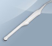
Конвексный датчик 4-9 МГц (ректо-вагинальный)
Акушерские исследования (ранние сроки), гинекология (матка, яичники), урология (предстательная железа), исследования прямой кишки.
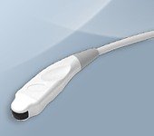
Линейные датчики
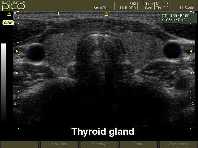
Линейный датчик 5-12 МГц
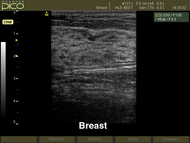
Поверхностные структуры (щитовидная железа, молочная железа, лимфоузлы), мускулоскелетные исследования (суставы, мышцы, подкожные структуры), периферические сосуды.
Линейный датчик 5-9 МГц
Поверхностные структуры (щитовидная железа, молочная железа, лимфоузлы), мускулоскелетные исследования (суставы, мышцы, подкожные структуры), периферические сосуды.
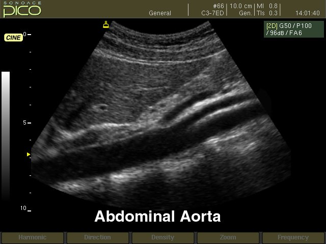
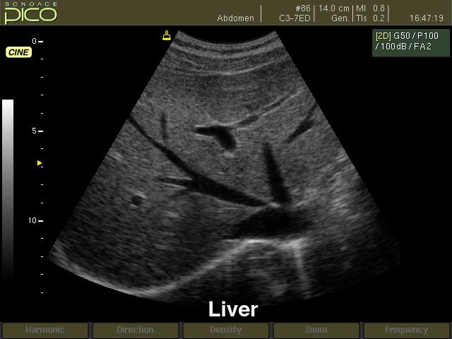
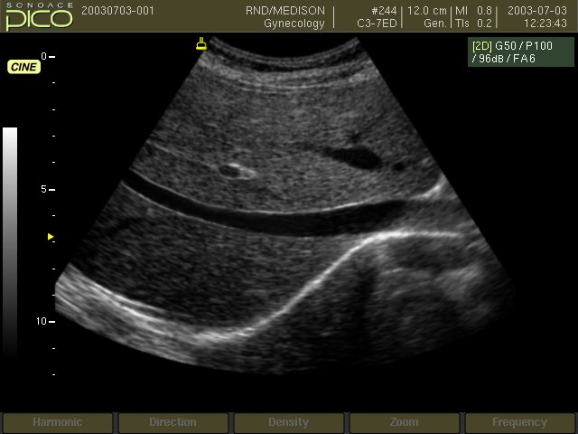
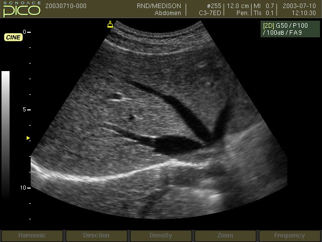
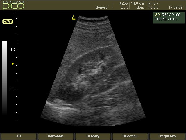
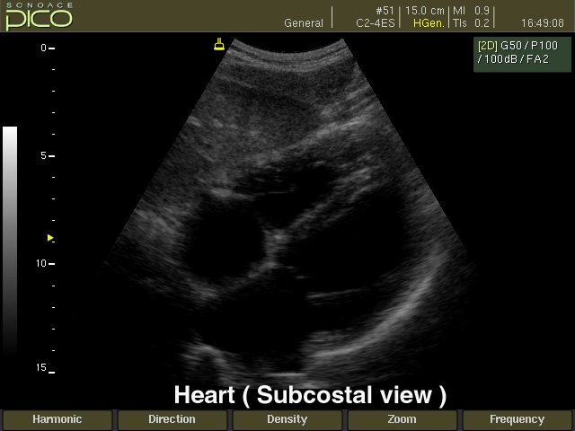
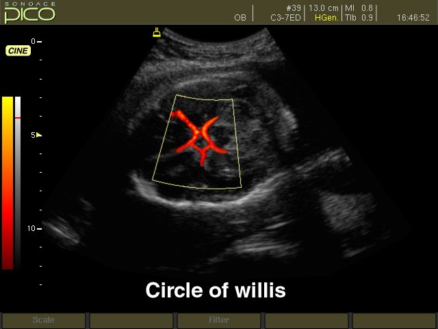
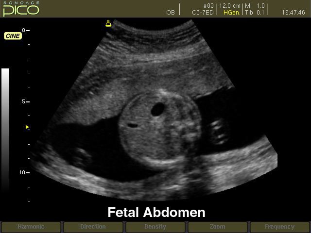
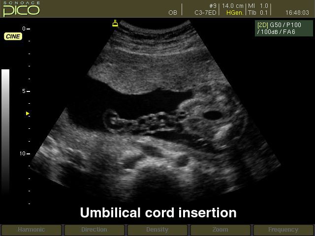
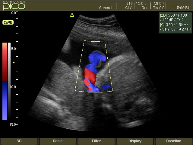
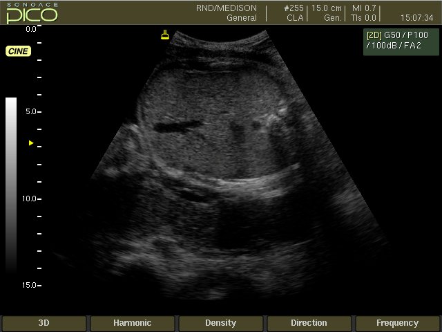
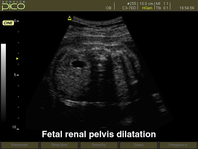
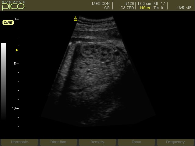
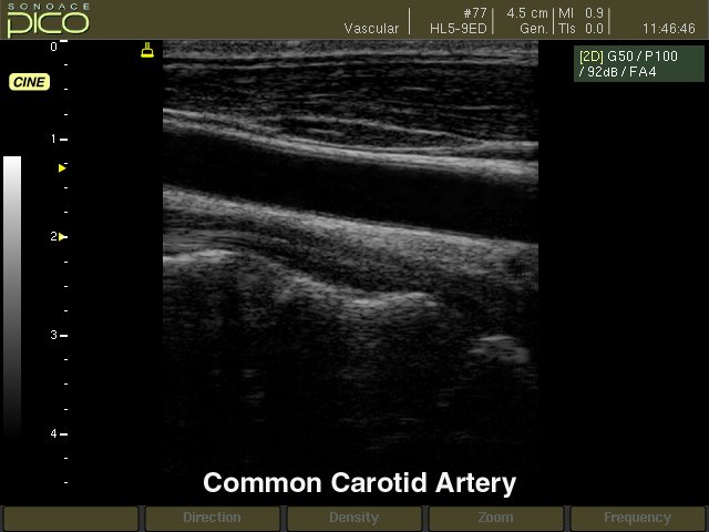
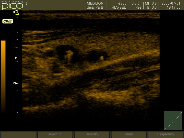
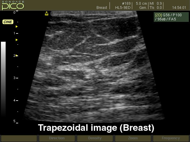
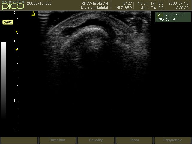
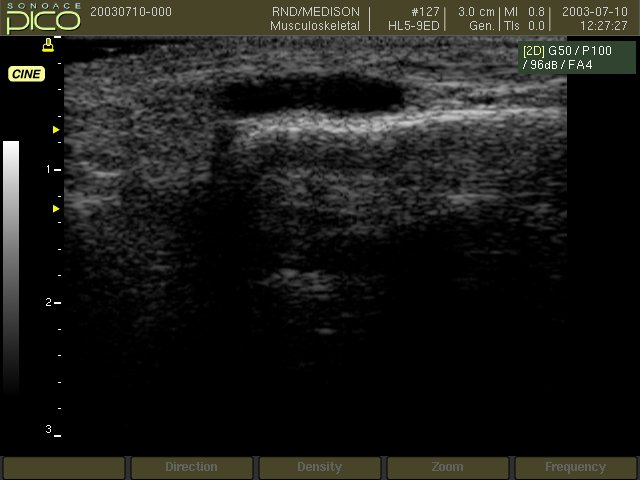
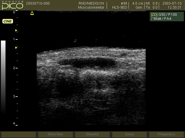
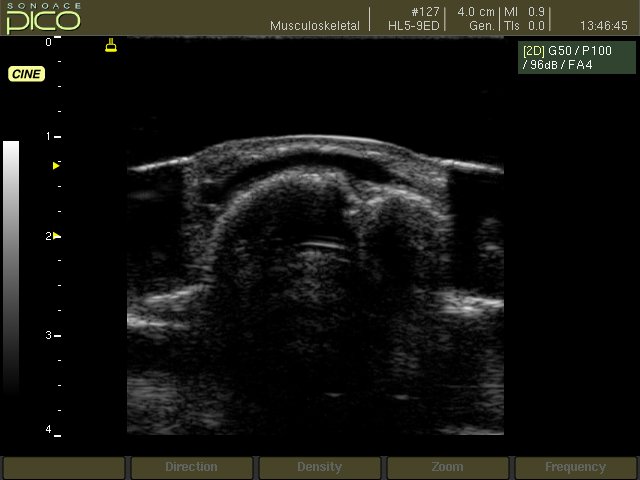
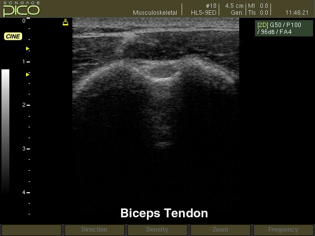
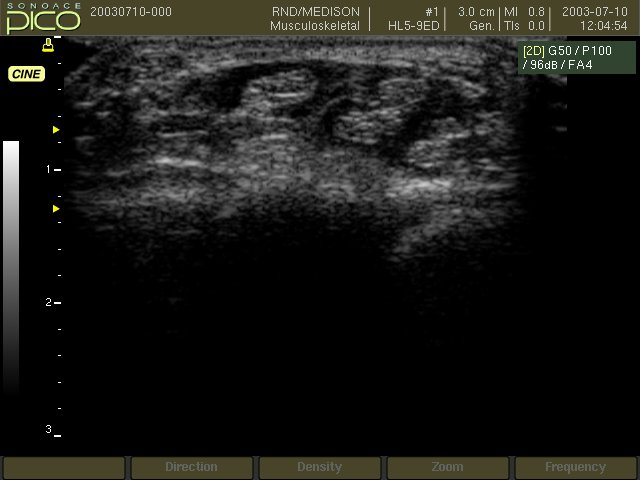
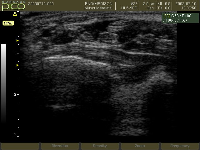
Примеры УЗИ на SonoAce-Pico (эхограммы)


Внимание! Копирование или частичная перепечатка материалов проспекта сканера "SonoAce Pico" на русском языке и размещение в интернет без согласия владельца (ЗАО "Медиэйс") запрещена, запрос на копирование материала можно отправить здесь.