УЗИ сканер SonoAce-8000 (Medison, снят с производства)
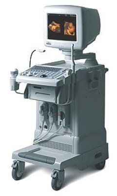
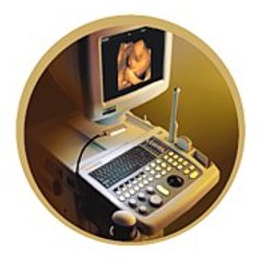
SonoAce-8000 Live - многофункциональный ультразвуковой сканер, 3D Live модификация аппарата SonoAce-8000 компании Medison с цветным, энергетическим, тканевым, импульсным и непрерывноволновым допплером, трехмерным УЗИ в реальном времени (4D объемными датчики).
Оптимальное сочетание "цена-качество" для современных диагностических центров, больниц и поликлиник, использующих в работе технологии трехмерного УЗИ, в частности трехмерное УЗИ плода.
Базовая комплектация: сканер SonoAce 8000 Live (монитор 15"; встроенные модули: цветного допплеровского картирования, энергетического допплера, импульсного допплера, 2-я гармоника, FreeHand 3D, Live 3D, SonoView-II; кинопамять; встроенная клавиатура с трекболом), флакон геля 250 мл и руководство оператора.
Опции для сканера SonoAce 8000 Live: кардиопакет: тканевый допплер (TDI) + цветной М-режим (CM) + программное обеспечение; непрерывноволновой допплер (CW); ЭКГ модуль; устройства хранения информации (USB флеш-карта, CD-RW); система DICOM.
Области применения: акушерство и гинекология, абдоминальные исследования и маммология, урология и эхокардиография, поверхностно расположенные органы и исследования сосудов, мускуло-скелетные исследования, а также педиатрия, неонаталогия, интраоперационные исследования, исследования с применением контрастных веществ.
Основные характеристики УЗ сканера SonoAce 8000 Live
- Стационарный ультразвуковой сканер.
- Монитор - 15" (36 см).
- Кардиопакет (опция).
- ЭКГ модуль (опция).
- Режимы сканирования: B, 2B, M, B+M;
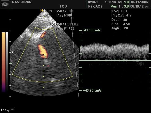
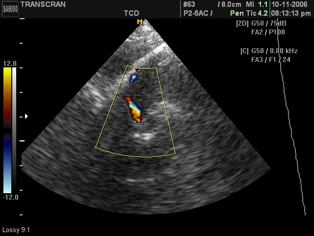
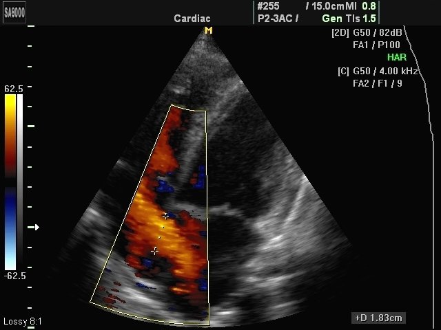
- CFM - цветное допплеровское картирование;
- PD - энергетический допплер (в т.ч. 3D);
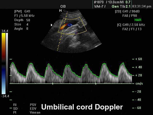
- PW - импульсный допплер;
- CW - непрерывноволновой допплер (опция). - Особенности сканирования:
- тканевая гармоника (регистрация 2-й гармоники эхосигнала, в том числе с помощью инверсной технологии);
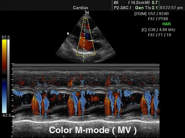
- цветной М-режим (опция);
- тканевый допплер (опция) - тканевая цветовая и спектральная допплерография для оценки сократительной способности миокарда;
- автоматический анализ допплеровских кривых;
- глубина сканирования до 30 см;
- steering - возможность изменения допплеровского угла в режимах CFM и PD;
- дуплексный и триплексный режим. - Разъемы для одновременного подключения до 3-х датчиков.
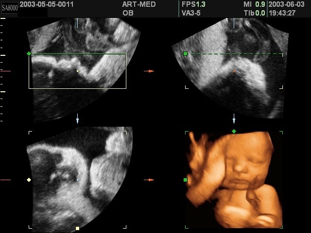
- Система FreeHand 3D - восстановление объемной структуры поверхностей тканей (функции увеличения, вращения и т.д.) при работе с обычными датчиками; восстановление трехмерной структуры сосудов в режиме энергетического допплера.
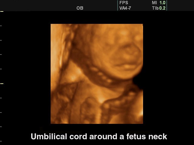
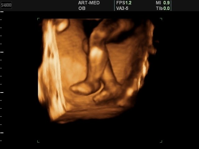
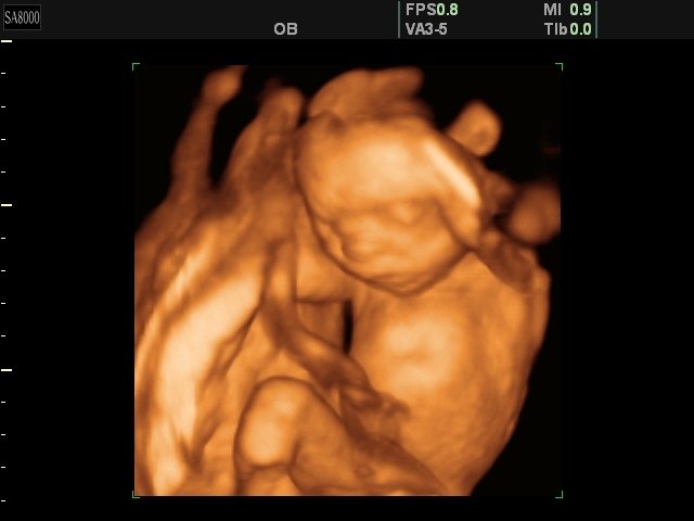
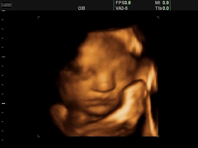
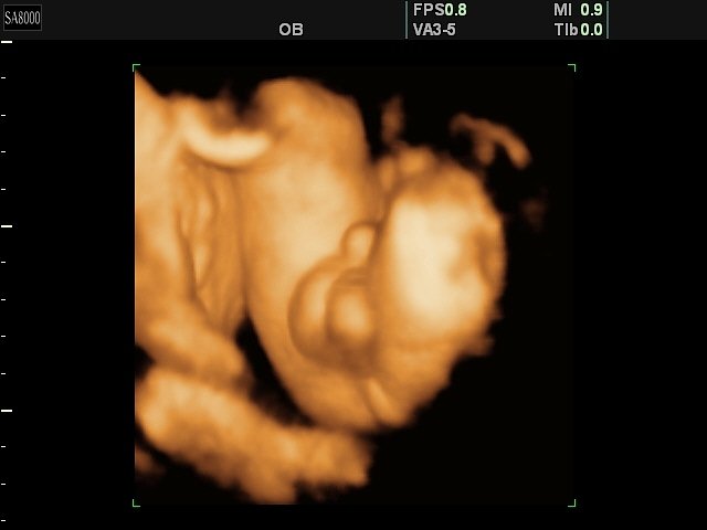
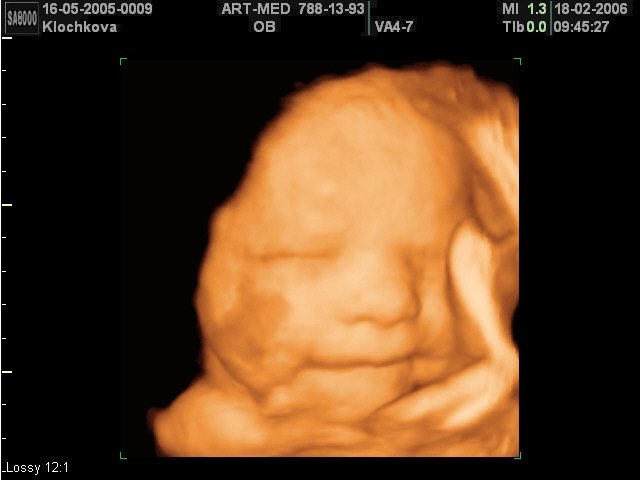
- Система Live 3D - возможность проведения трехмерного УЗИ в реальном времени (4D):
- 3D датчики;
- получение любого среза в каждой из 3-х проекций;
- получение трехмерных изображений в режиме серой шкалы, а также цветного и энергетического допплера;
- кинопетля в 3D режиме;
- фото-режим;
- измерения в трехмерном режиме;
- функция автоматического вычисления объема структур сложной формы. - SonoAtlas - программа обучения проведению ультразвуковой диагностики (электронный учебник с примерами эхограмм и описанием методики их получения).
- Система SonoView - система архивации и дальнейшего просмотра статических и динамических изображений. Имеется возможность проведения измерений в архиве. При наличии соответствующих приводов возможно копирование изображений на гибкие дискеты, компакт-диски, магнитооптику.
- Система DICOM (опция) - возможность сетевой интеграции с PACS-системами (например, для архивации или печати ультразвуковых эхограмм на оборудовании других производителей медтехники).
Инновационные технологии
- Multi-beam, Optimal Volume Resolution, Optimum Tissue Imaging, Tissue Harmonic Imaging, Optimized Harmonic Imaging, Pulse Inversion Harmonic, FINE, CAFE.
- VOCAL (Virtual Organ Computer Aided anaLysis) - программа вычисления объемов структур сложной формы в трехмерном режиме. Основана на алгоритме автоматического обозначения контуров структур при трехмерной реконструкции, что позволяет с максимальной точностью вычислить объем структур любой формы (предстательная железа, кисты и т.д.).
- See-Thru - технология, использующая объединение трехмерного энергетического допплера и серошкального изображения для улучшения визуализации сосудов в области патологии (опухоли).
- 3D image optimizing - режимы акустической прозрачности трехмерного ультразвукового изображения (Surface mode, Maximum mode, Minimum mode, X-Ray mode).
Пакеты ультразвуковых диагностических программ
- Основные измерения: измерения расстояния, окружности, площади, объема; измерение тазобедренного сустава; измерение расстояния в M-режиме; измерение скорости в спектральном допплеровском режиме и др.
- Пакет гинекологических исследований: матка, левый и правый яичники, левая и правая почки, артерии левого и правого яичников, левый и правый фолликулы.
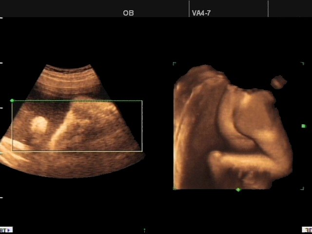
- Пакет акушерских исследований: биометрия плода, краниологическое исследование плода, исследование длинных костей плода, измерение индекса околоплодных вод (AFI), допплер плода и др.
- Биометрия плода включает измерения теменно-копчиковой длинны (CRL), размера плодного пузыря (GS), бипариетальный размера головки плода (BPD), затылочно-лобного расстояния (OFD), длины окружности головы плода (НC), передне-заднего размера брюшной полости (APD), поперечного размера брюшной полости (TAD), окружности живота (AC), площади сечения тела (FTA), длины бедра (FL), поперечного (TTD) и передне-заднего (APTD) размеров тела плода.
- Краниологическое исследование плода включает измерения параметров мозжечка (CEREB), а также внешнего (OOD) и внутреннего (IOD) межглазных расстояний.
- Исследование длинных костей плода включает измерения длины плечевой кости (Humerus), локтевой кости (Ulna), большеберцовой кости (Tibia), лучевой кости (Rad), ключицы (Clav) и позвоночника (LV).
Кроме того, семь уравнений для оценки веса плода: Хедлок (Hadlock) 1-4, Хансман (Hansmann) и Мерц (Merz); ЧСС плода (Fetal HR); таблицы, определяемые пользователем. - Пакет урологических расчетов: разностный объем, объем предстатательной железы, вычисление плотности простатспецифического антигена (PSA).
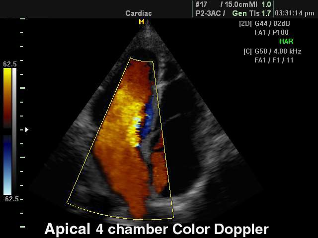
- Пакет кардиологических исследований:
- в 2D-режиме рассчитываются значения таких параметров, как объем по методу Симпсона (Simpson), объем по площади и длине, двумерные характериcтики (например, фракция выброса левого желудочка) и масса левого желудочка;
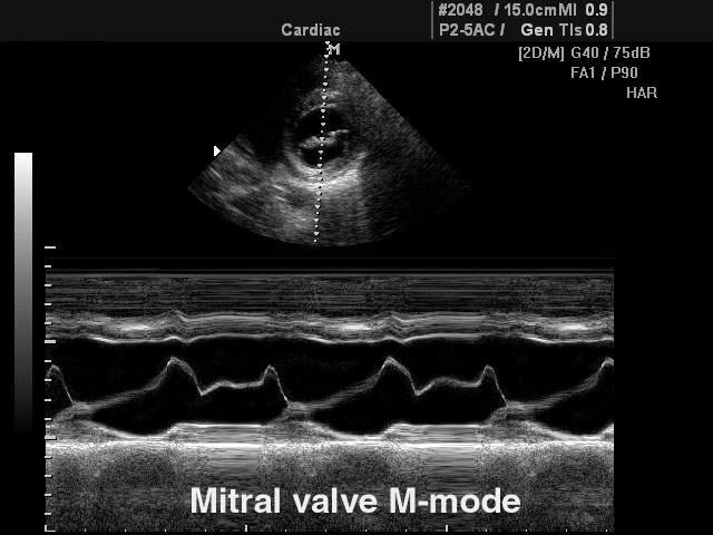
- в M-режиме вычисляются значения параметров для левого желудочка, аорты и левого предсердия, митрального клапана, а также частота сердечных сокращений. - Пакет расчетов параметров сосудов: вычисления объемного кровотока, процента стеноза, индекса сопротивления (RI), пульсационного индекса (PI) и др.
Датчики для сканера SonoAce 8000 Live
Конвексные датчики
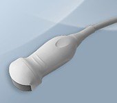
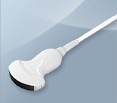
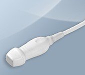
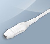
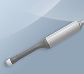
Конвексный датчик C 2-4ES/20/120 (микроконвексный)
Кардиология, абдоминальные исследования (печень, желчный пузырь, поджелудочная железа, селезенка, глубокие сосуды), почки.
Биопсийный набор: нет.
Конвексный датчик C 2-5EL/40/85
Акушерские исследования (плод, сердце плода), гинекология (матка, яичники), абдоминальные исследования (печень, желчный пузырь, поджелудочная железа, селезенка, глубокие сосуды), почки.
Биопсийный набор: есть.
Конвексный датчик C 2-5ET/40/76
Акушерские исследования (плод, сердце плода), гинекология (матка, яичники), абдоминальные исследования (печень, желчный пузырь, поджелудочная железа, селезенка, глубокие сосуды), почки.
Биопсийный набор: есть.
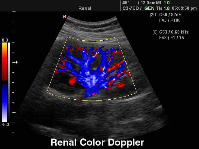
Конвексный датчик C 3-7ED/50/70
Акушерские исследования (плод, сердце плода), гинекология (матка, яичники), абдоминальные исследования (печень, желчный пузырь, поджелудочная железа, селезенка, глубокие сосуды), почки.
Биопсийный набор: есть.
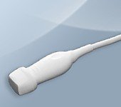
Конвексный датчик C 4-9ED/10/150 (микроконвексный)
Неонатология и педиатрия: абдоминальные исследования, почки, сердце, глубокие сосуды, мозг.
Биопсийный набор: нет.
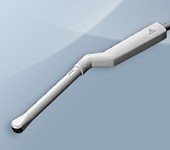
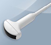
Конвексный датчик EC 4-9ED/10/150 (вагинальный)
Акушерские исследования (ранние сроки), гинекология (матка, яичники), урология (предстательная железа), исследования прямой кишки.
Биопсийный набор: есть.
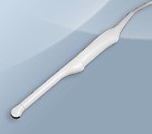
Конвексный датчик EC 4-9ES/10/150 (ректо-вагинальный)
Акушерские исследования (ранние сроки), гинекология (матка, яичники), урология (предстательная железа), исследования прямой кишки.
Биопсийный набор: есть.
Фазированные датчики
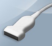
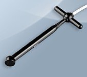
Фазированный датчик P 2-5AC/15
Кардиология и транскраниальные исследования у взрослых.
Биопсийный набор: нет.
Фазированный датчик P 2-5AC/19
Кардиология и транскраниальные исследования у взрослых.
Биопсийный набор: нет.
Фазированный датчик P 3-7AC/10
Кардиология и транскраниальные исследования у детей.
Биопсийный набор: нет.
Линейные датчики
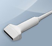
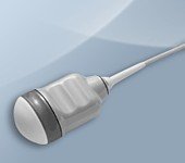
Линейный датчик HL 5-12ED/40
Поверхностные структуры (щитовидная железа, молочная железа, лимфоузлы), мускулоскелетные исследования (суставы, мышцы, подкожные структуры), периферические сосуды.
Биопсийный набор: есть.
Линейный датчик L 5-9EC/40
Поверхностные структуры (щитовидная железа, молочная железа, лимфоузлы), мускулоскелетные исследования (суставы, мышцы, подкожные структуры), периферические сосуды.
Биопсийный набор: есть.
Линейный датчик L 5-9ER/50
Поверхностные структуры (щитовидная железа, молочная железа, лимфоузлы), мускулоскелетные исследования (суставы, мышцы, подкожные структуры), периферические сосуды.
Биопсийный набор: есть.
Объемные датчики

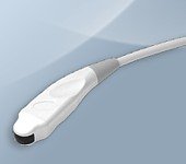
Объемный датчик 3D 3-5EK/40/69
Трехмерные абдоминальные исследования, акушерство и гинекология.
Биопсийный набор: есть.

Объемный датчик 3D 4-8ET/40/84
Трехмерные абдоминальные исследования, акушерство (трехмерное УЗИ плода) и гинекология.
Биопсийный набор: есть.
Объемный датчик 3D 5-8EK/10/128 (ректо-вагинальный)
Трехмерные исследования в акушерстве (ранние сроки), гинекологии (матка, яичники), урологии (предстательная железа), исследования прямой кишки.
Биопсийный набор: есть.
Объемный датчик VAW 4-7/40/69
Трехмерные абдоминальные исследования, акушерство (трехмерное УЗИ плода) и гинекология.
Биопсийный набор: есть.

Объемный датчик VDW 5-8/10/128 (ректо-вагинальный)
Трехмерные исследования в акушерстве (ранние сроки), гинекологии (матка, яичники), урологии (предстательная железа), исследования прямой кишки.
Биопсийный набор: есть.
Допплеровские датчики
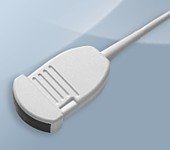
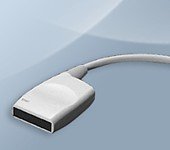
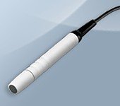
Допплеровский датчик CW 2.0 (слепой допплер)
Транскраниальные исследования, сосуды.
Биопсийный набор: нет.
Допплеровский датчик CW 4.0 (слепой допплер)
Транскраниальные исследования, сосуды.
Биопсийный набор: нет.
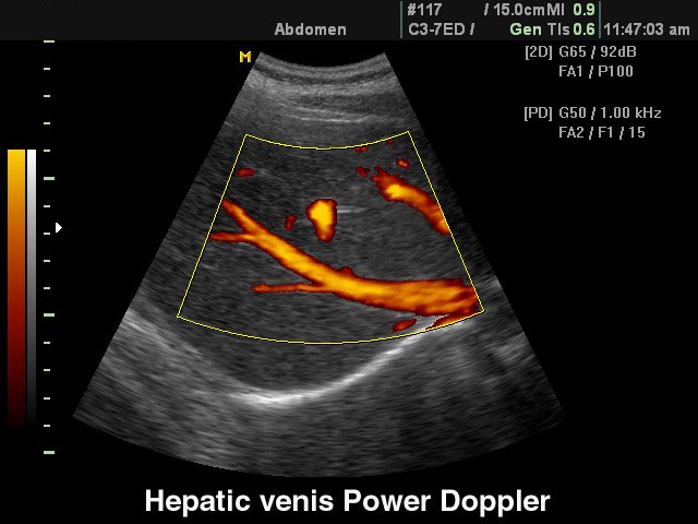
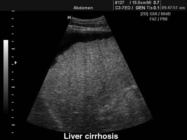
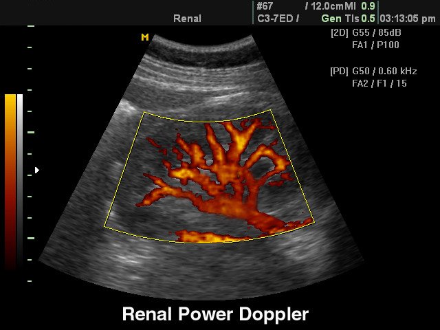
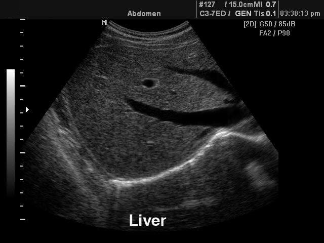
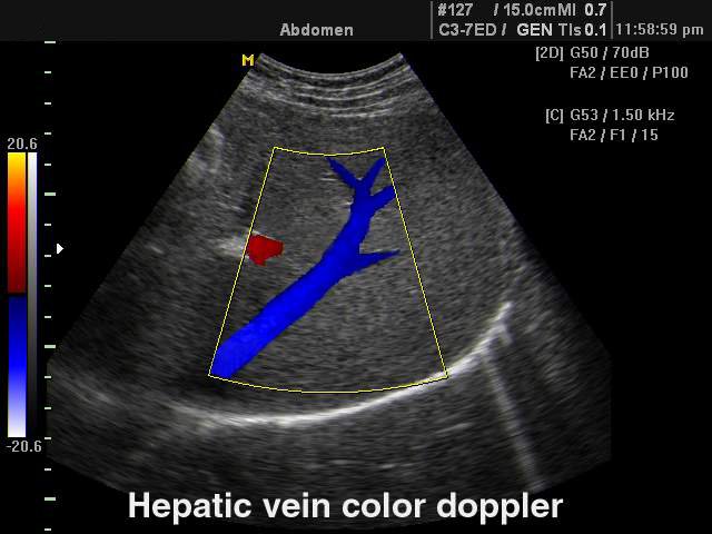
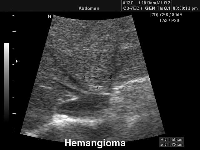
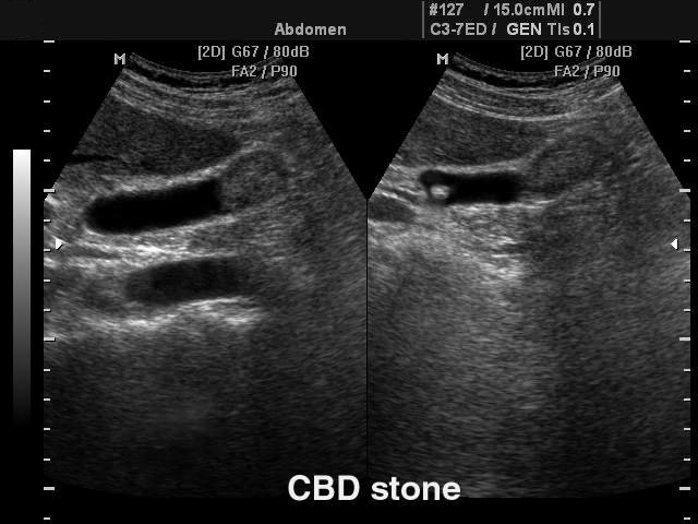
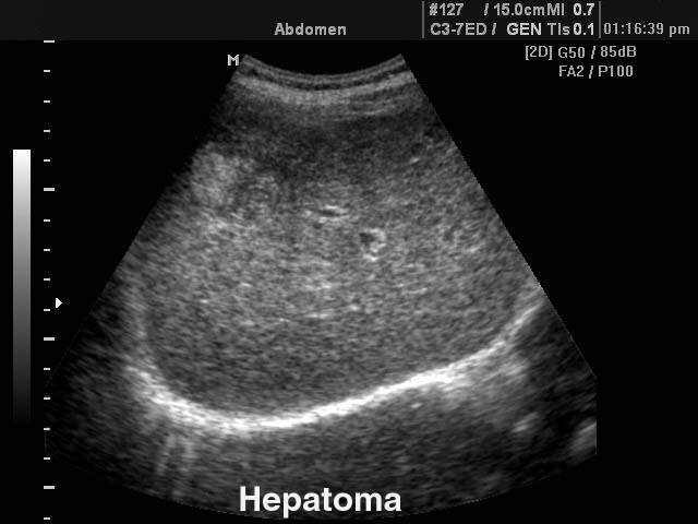
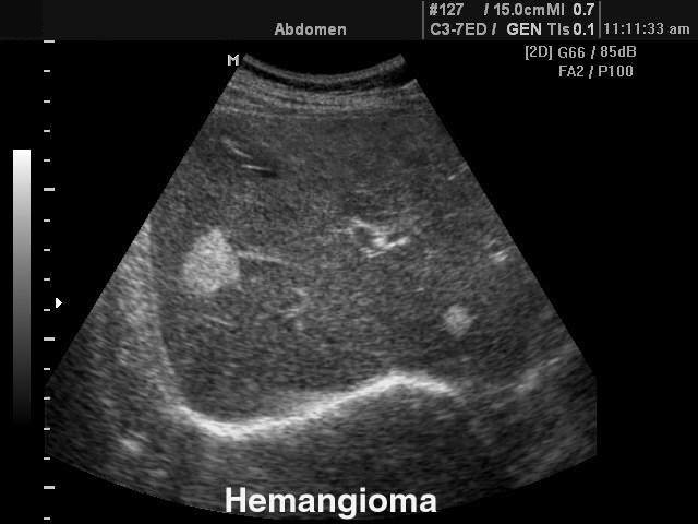
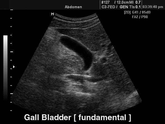
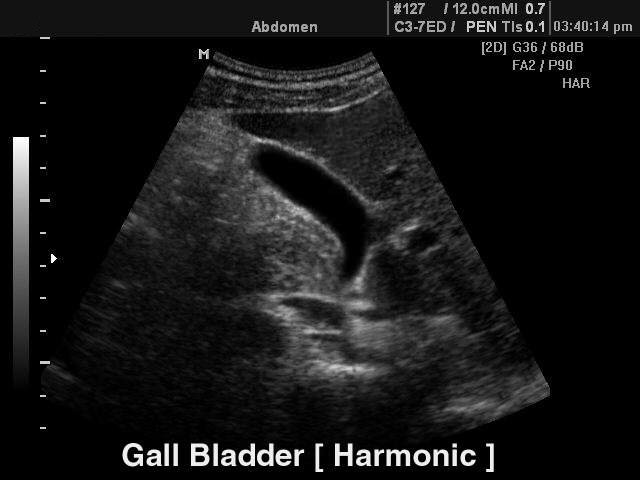
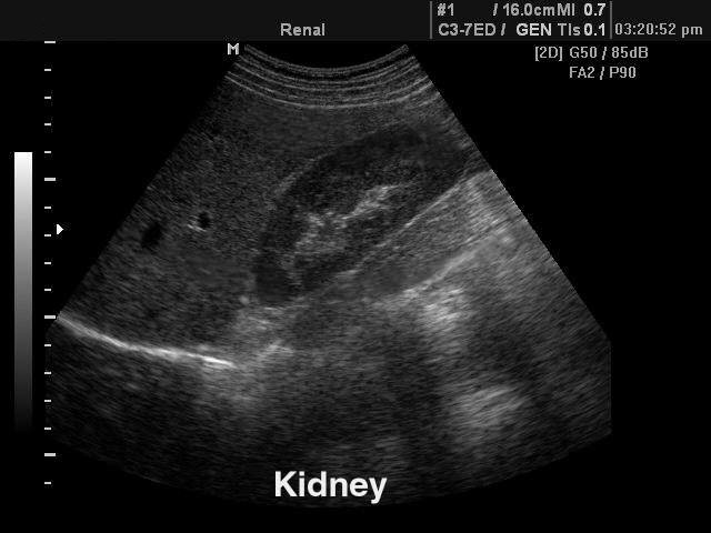
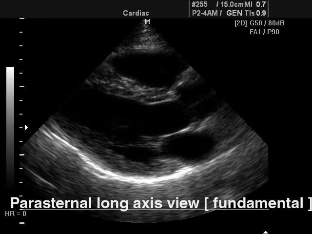
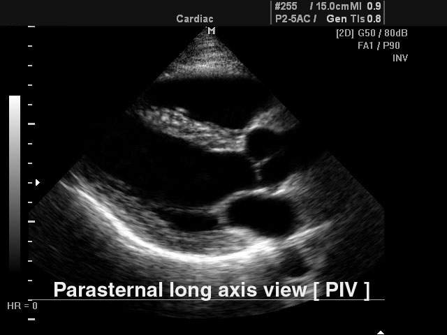
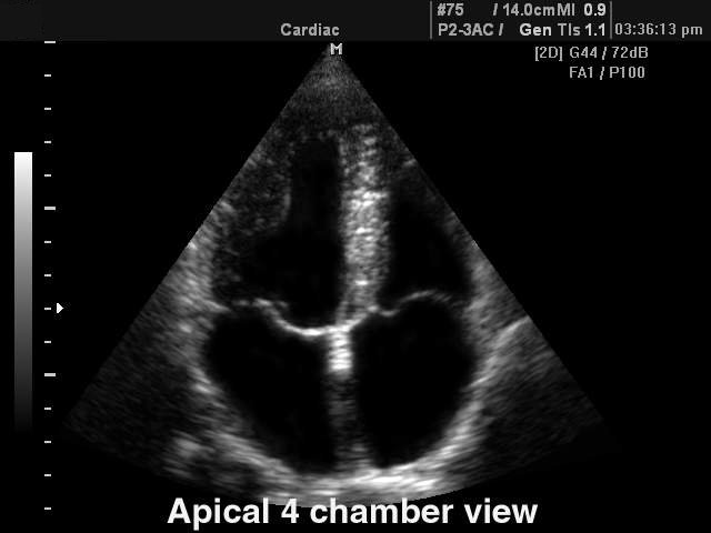
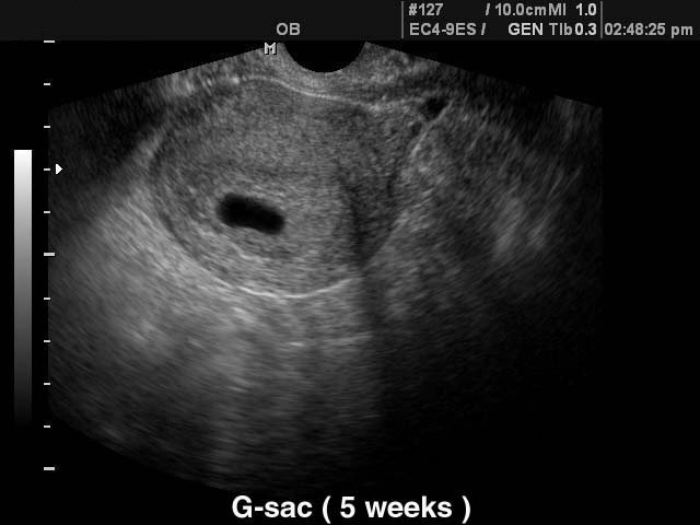
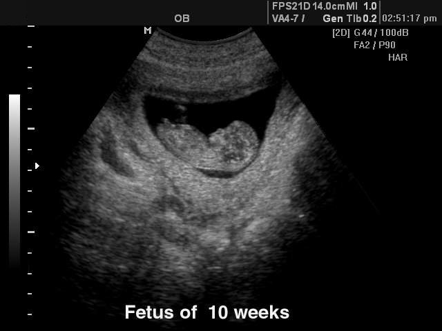
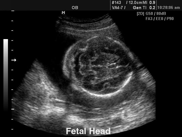
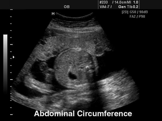
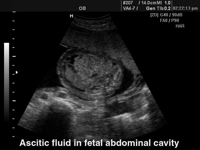
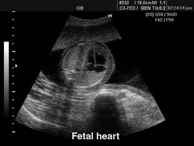
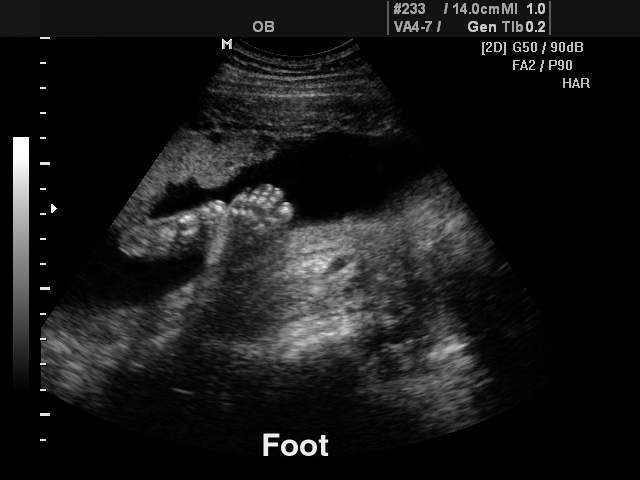
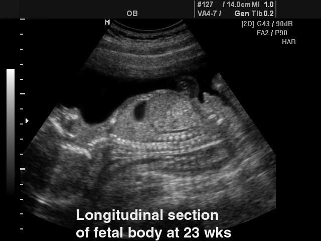
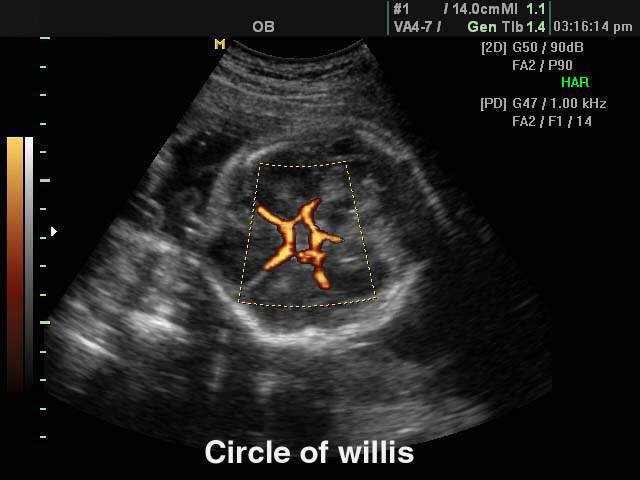
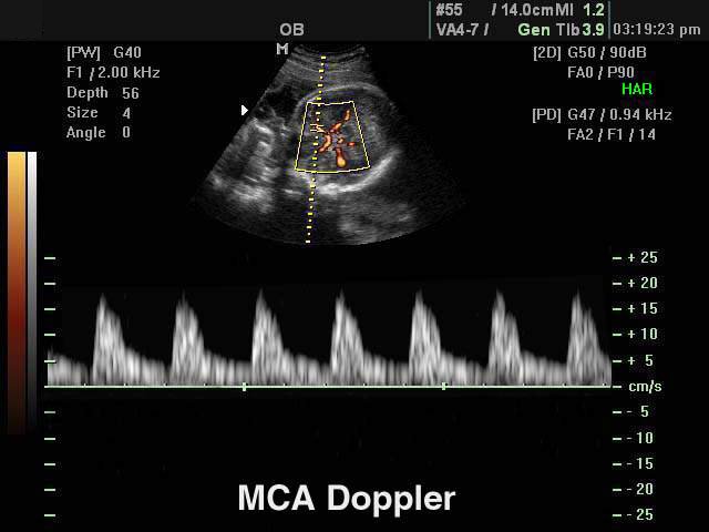
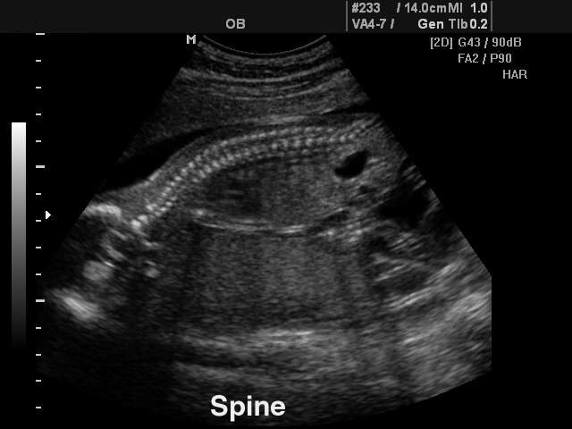
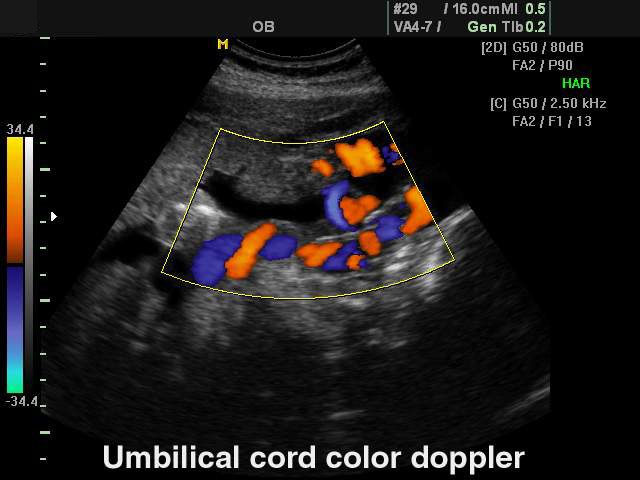
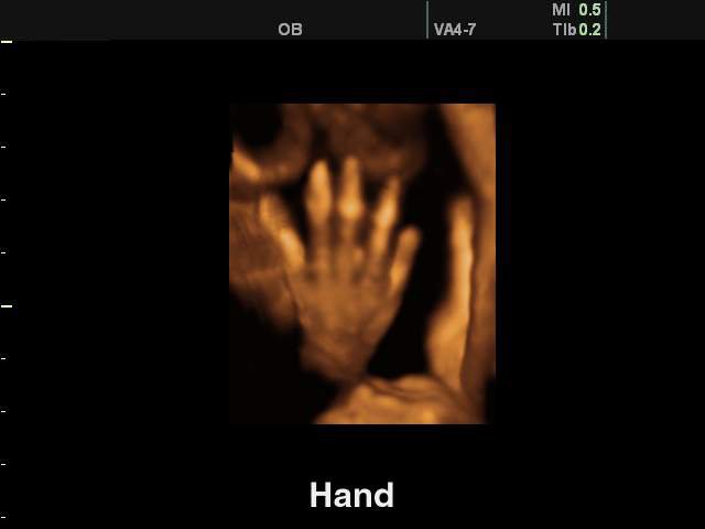
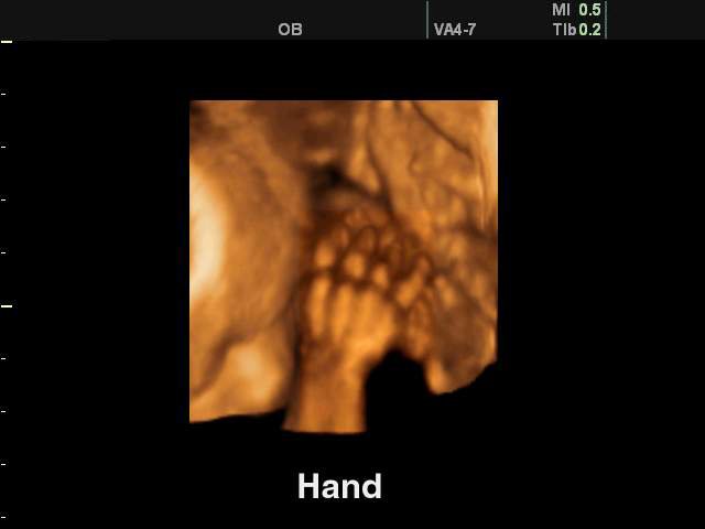
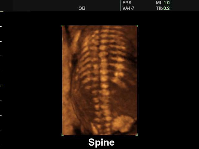
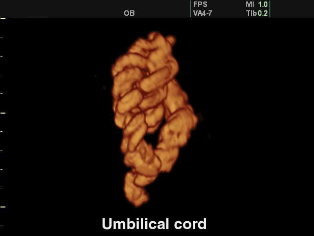
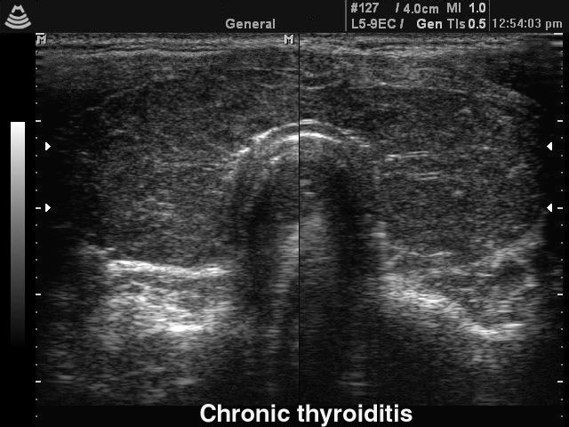
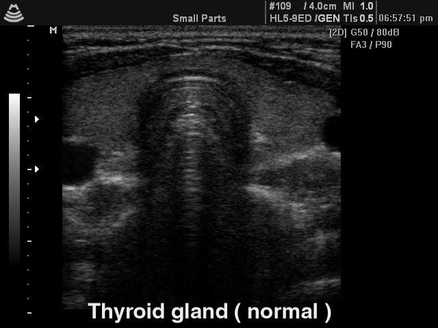
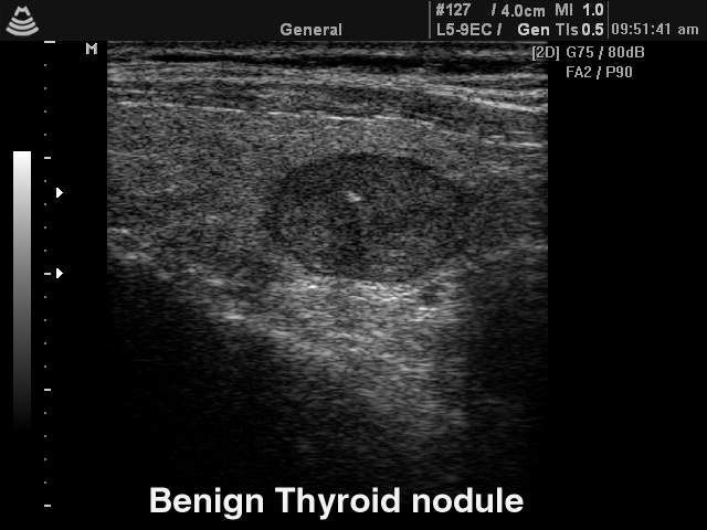
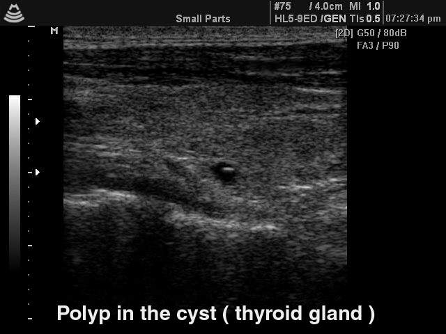
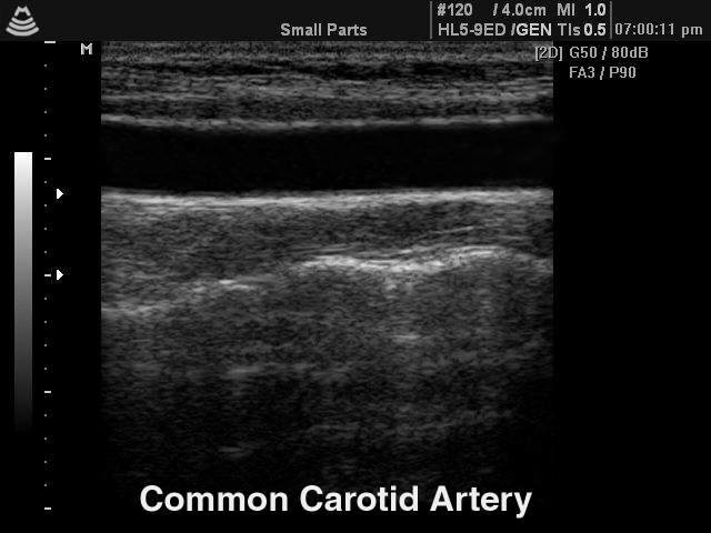
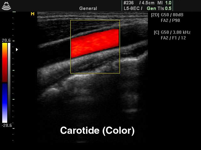
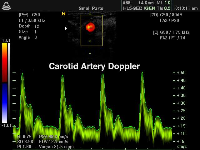
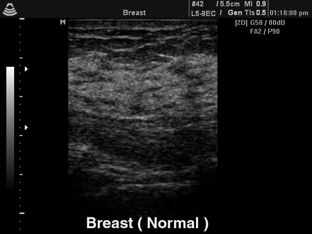
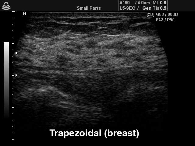
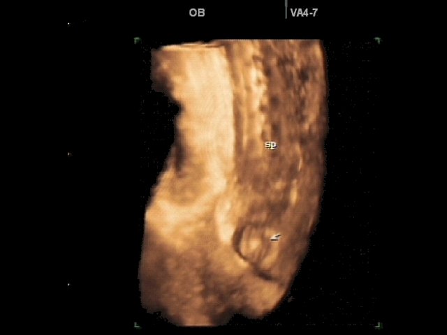
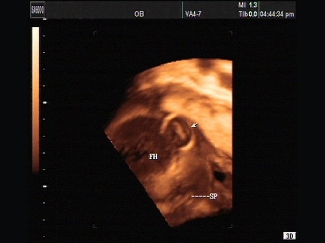
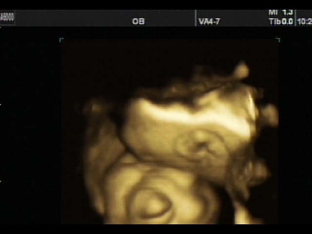
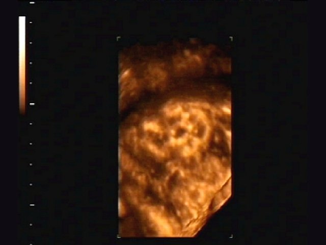
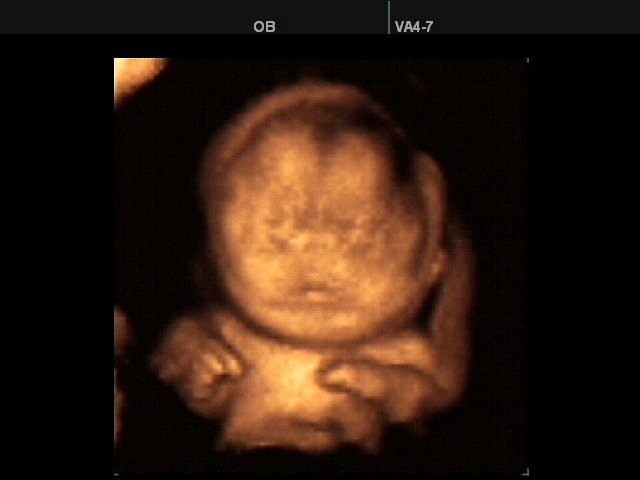
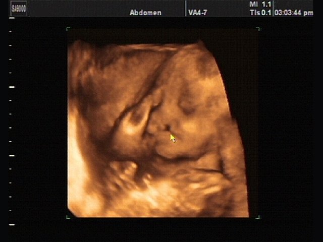
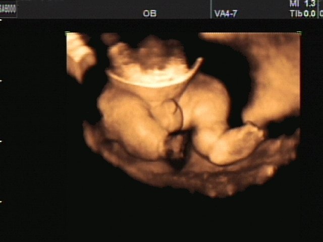
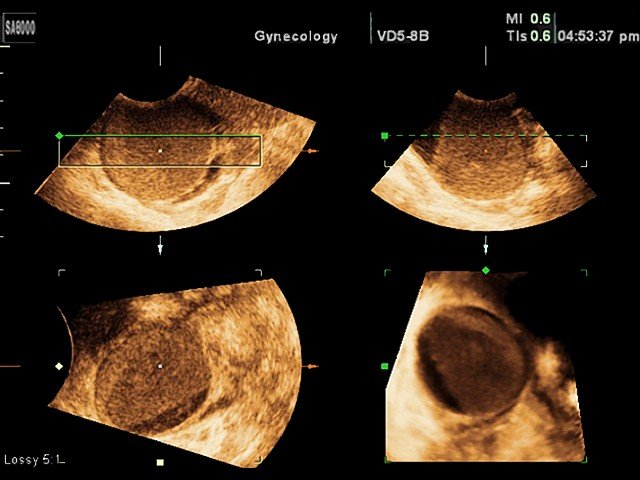
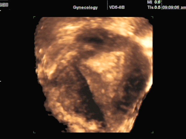
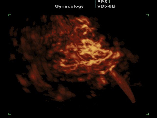
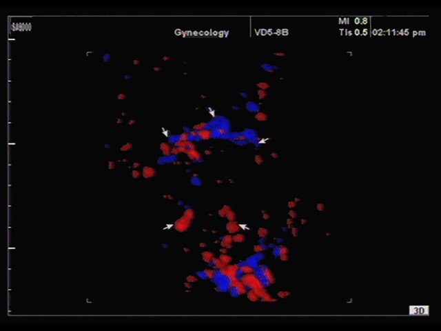
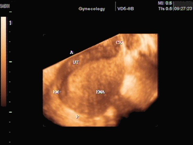
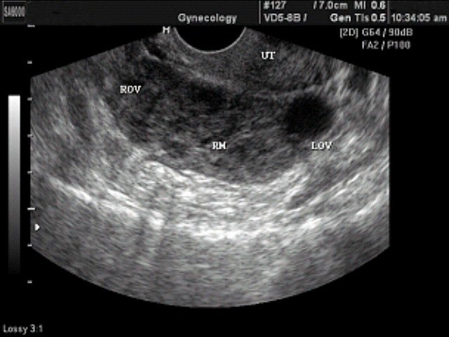
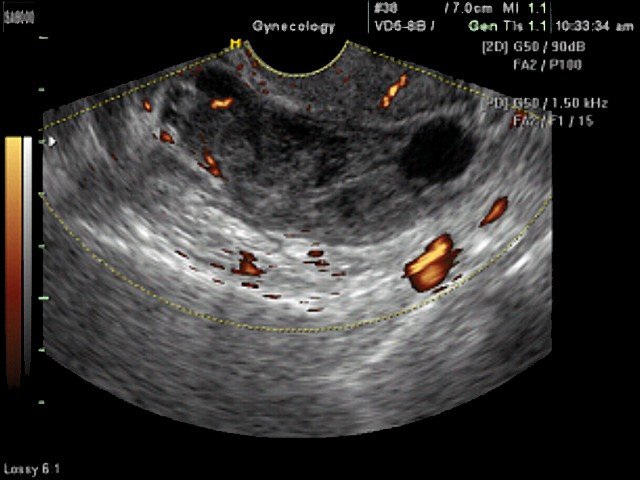
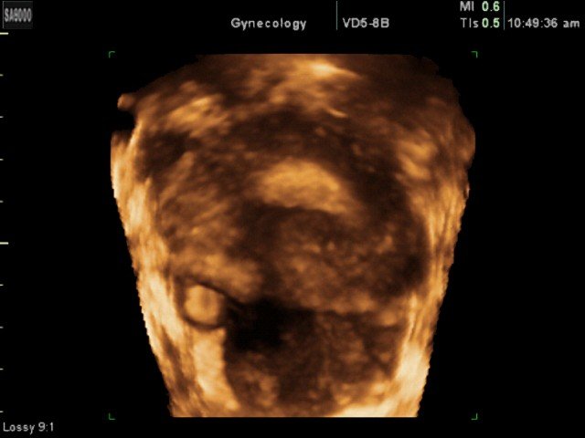
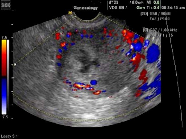
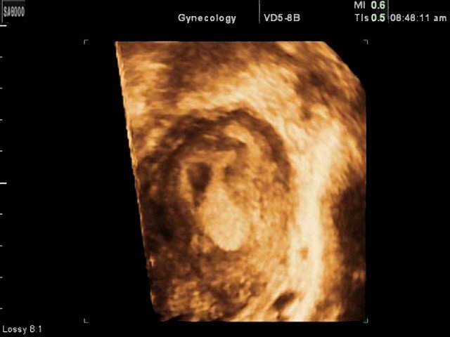
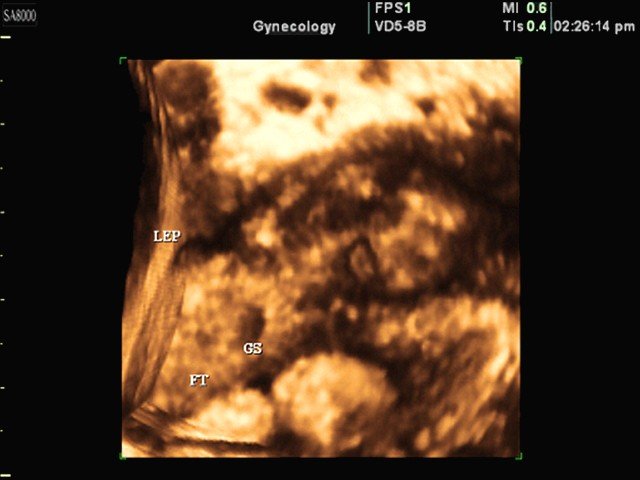
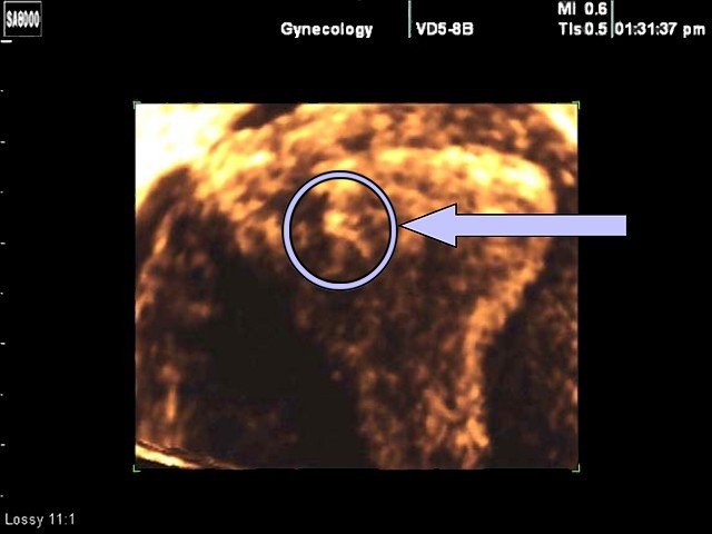
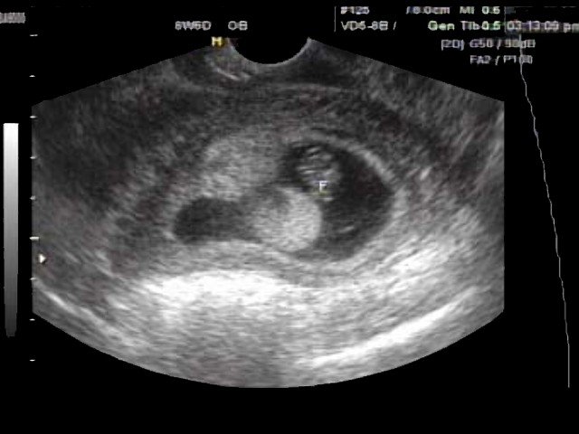
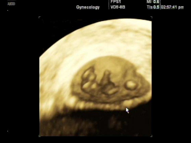
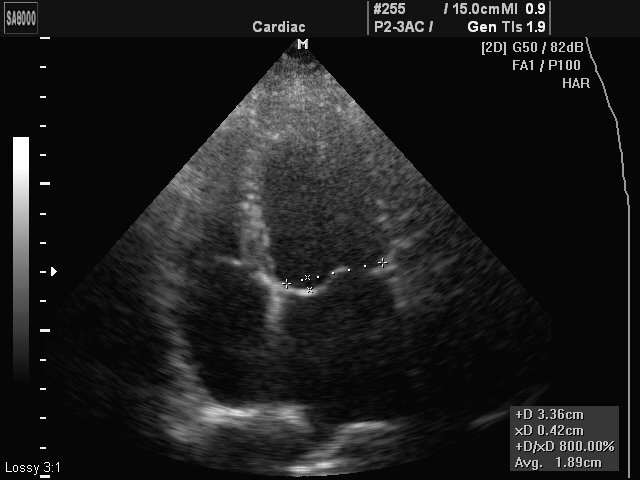
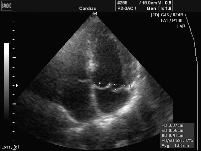
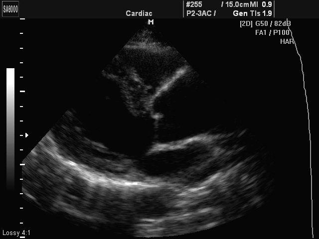
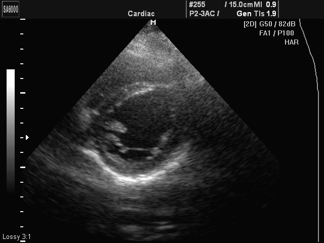
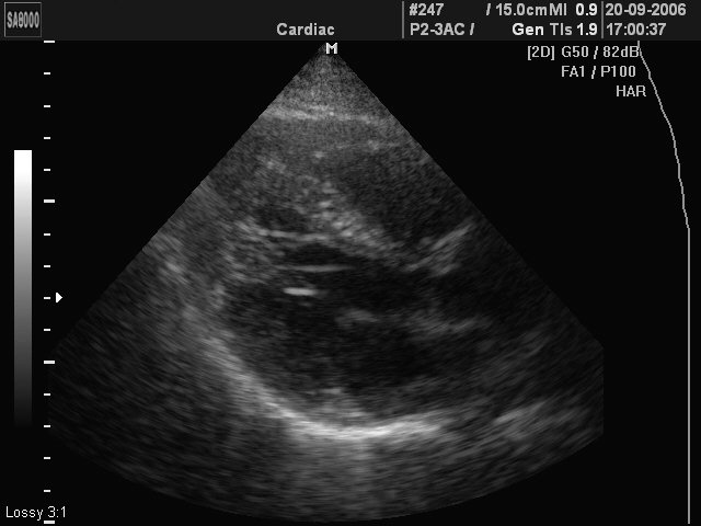
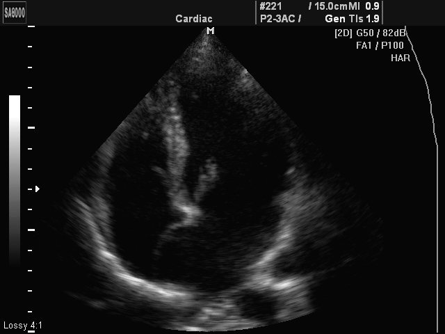
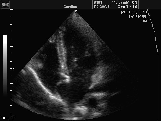
Примеры УЗИ на сканере SonoAce 8000 Live (эхограммы)
Внимание! Копирование или частичная перепечатка материалов проспекта сканера "SonoAce 8000 Live" на русском языке и размещение в интернет без согласия владельца (ЗАО "Медиэйс") запрещена, запрос на копирование материала можно отправить здесь.