УЗИ сканер SonoAce-9900 (Medison, снят с производства)
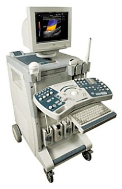
SonoAce-9900 - ультразвуковой сканер компании Medison с цветовым допплеровским картированием, энергетическим, импульсным, тканевым и непрерывноволновым допплером, трехмерное УЗИ в реальном времени (3D обычными и 4D объемными датчиками).
Оптимальное решение для современных диагностических центров, медицинских исследовательских институтов, использующих в работе технологии трехмерного УЗИ и стресс-эхо в кардиологии.
Базовая комплектация: сканер SonoAce 9900 (монитор 15"; встроенные модули: цветного допплеровского картирования, энергетического допплера, импульсного допплера, 2-я гармоника, FreeHand 3D, SonoView-II, кинопамять; встроенный дисковод: CD-RW; встроенная клавиатура с трекболом), флакон геля 250 мл и руководство оператора.
Опции для сканера SonoAce 9900: cистема Live 3D; кардио-система (ЭКГ модуль + программное обеспечение); непрерывноволновой допплер; цветной М-режим; тканевый допплер; стресс-эхо; устройства хранения информации (USB флеш-карта, магнитооптика, DVD-RW); система DICOM.
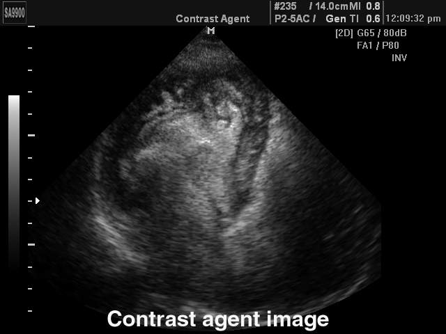
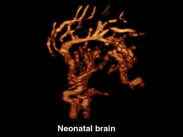
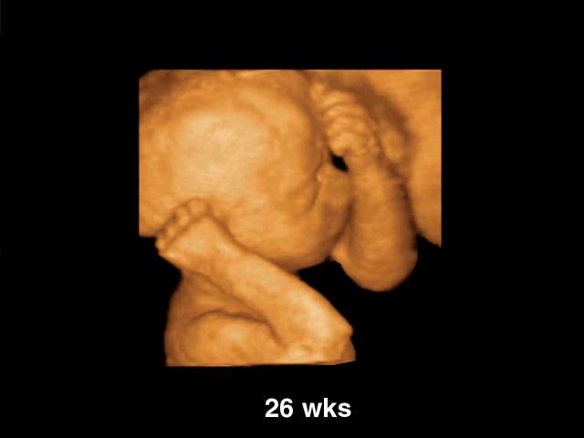
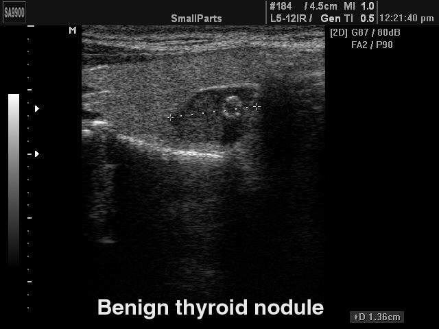
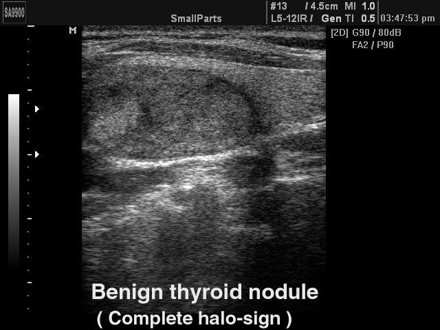
Области применения: акушерство и гинекология, абдоминальные исследования и маммология, урология и эхокардиография, поверхностно расположенные органы и исследования сосудов, мускуло-скелетные исследования, а также педиатрия, неонаталогия, транскраниальные исследования, интраоперационные исследования, исследования с применением контрастных веществ.
Основные характеристики УЗ сканера SonoAce-9900
- Стационарный ультразвуковой сканер.
- Монитор - 15" (36 см).
- Кардио-система: ЭКГ модуль + программное обеспечение (опция).
- Режимы сканирования: B, 2B, M, B+M;
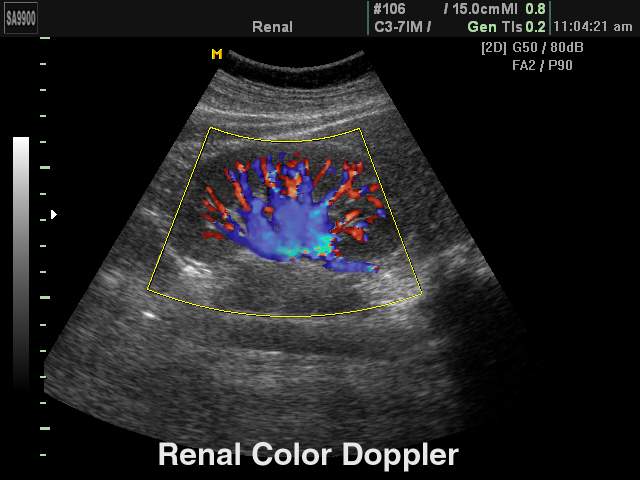
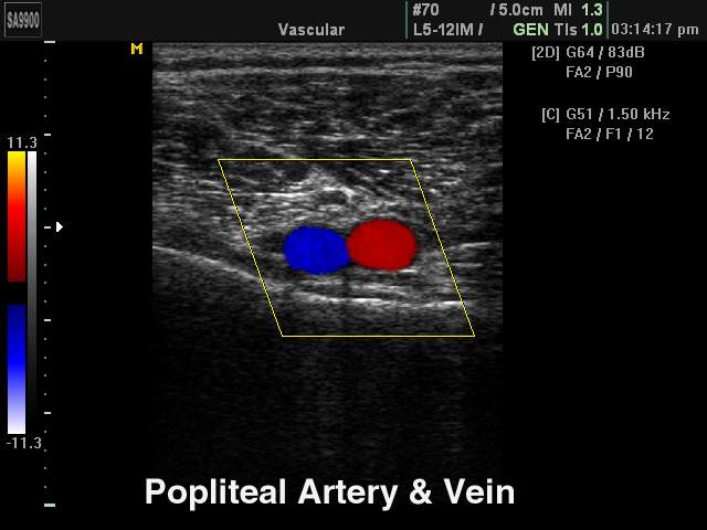
- CFM - цветное допплеровское картирование;
- PD - энергетический допплер (в т.ч. 3D);
- PW - импульсный допплер;
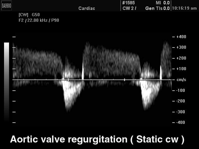
- CW - непрерывноволновой допплер (опция). - Особенности сканирования:
- тканевая гармоника (регистрация 2-й гармоники эхосигнала, в том числе с помощью инверсной технологии);
- цветной М-режим (опция);
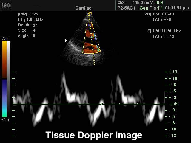
- тканевый допплер - тканевая цветовая и спектральная допплерография для оценки сократительной способности миокарда (опция);
- автоматический анализ допплеровских кривых;
- глубина сканирования до 30 см;
- steering - возможность изменения допплеровского угла в режимах CFM и PD;
- дуплексный и триплексный режим;
- стресс-эхо - программа диагностики сердца (опция). - Разьемы для одновременного подключения до 4-х датчиков.
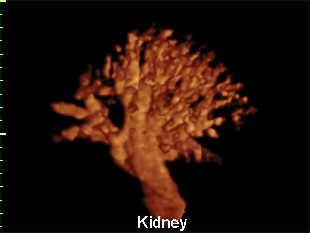
- Система FreeHand 3D - восстановление объемной структуры поверхностей тканей (функции увеличения, вращения и т.д.) при работе с обычными датчиками; восстановление объемной структуры сосудов в режиме энергетического допплера.
- Система Live 3D - возможность проведения трехмерного УЗИ в реальном времени (4D):
- 3D датчики;
- получение любого среза в каждой из 3-х проекций;
- получение трехмерных изображений в режиме серой шкалы, а также цветного и энергетического допплера.
- кинопетля в 3D режиме;
- фото-режим;
- измерения в объемном режиме;
- функция автоматического вычисления объема структур сложной формы. - Система SonoView - система архивации и дальнейшего просмотра статических и динамических изображений. Имеется возможность проведения измерений в архиве. При наличии соответствующих приводов возможно копирование изображений на гибкие дискеты, компакт-диски, магнитооптику.
- Система DICOM (опция) - возможность сетевой интеграции с PACS-системами (например, для архивации или печати ультразвуковых эхограмм на оборудовании других производителей медтехники).
Инновационные технологии
- Multi-beam, Optimal Volume Resolution, Optimum Tissue Imaging, Tissue Harmonic Imaging, Optimized Harmonic Imaging, Pulse Inversion Harmonic, FINE, CAFE.
- VOCAL (Virtual Organ Computer Aided anaLysis) - программа вычисления объемов структур сложной формы в трехмерном режиме. Основана на алгоритме автоматического обозначения контуров структур при трехмерной реконструкции, что позволяет с максимальной точностью вычислить объем структур любой формы (предстательная железа, кисты и т.д.).
- See-Thru - технология, использующая объединение трехмерного энергетического допплера и серошкального изображения для улучшения визуализации сосудов в области патологии (опухоли).
- 3D image optimizing - режимы акустической прозрачности трехмерного ультразвукового изображения (Surface mode, Maximum mode, Minimum mode, X-Ray mode).
Пакеты ультразвуковых диагностических программ
- Основные измерения: измерения расстояния, окружности, площади, объема; измерение тазобедренного сустава; измерение расстояния в M-режиме; измерение скорости в спектральном допплеровском режиме и др.
- Пакет гинекологических исследований: матка, левый и правый яичники, левая и правая почки, артерии левого и правого яичников, левый и правый фолликулы.
- Пакет акушерских исследований: биометрия плода, краниологическое исследование плода, исследование длинных костей плода, измерение индекса околоплодных вод (AFI), допплер плода и др. Биометрия плода включает измерения теменно-копчиковой длинны (CRL), размера плодного пузыря (GS), бипариетальный размера головки плода (BPD), затылочно-лобного расстояния (OFD), длины окружности головы плода (НC), передне-заднего размера брюшной полости (APD), поперечного размера брюшной полости (TAD), окружности живота (AC), площади сечения тела (FTA), длины бедра (FL), поперечного (TTD) и передне-заднего (APTD) размеров тела плода. Краниологическое исследование плода включает измерения параметров мозжечка (CEREB), а также внешнего (OOD) и внутреннего (IOD) межглазных расстояний. Исследование длинных костей плода включает измерения длины плечевой кости (Humerus), локтевой кости (Ulna), большеберцовой кости (Tibia), лучевой кости (Rad), ключицы (Clav) и позвоночника (LV).
Кроме того, семь уравнений для оценки веса плода: Хедлок (Hadlock) 1-4, Хансман (Hansmann) и Мерц (Merz); ЧСС плода (Fetal HR); таблицы, определяемые пользователем. - Пакет урологических расчетов: разностный объем, объем предстательной железы, вычисление плотности простатспецифического антигена (PSA).
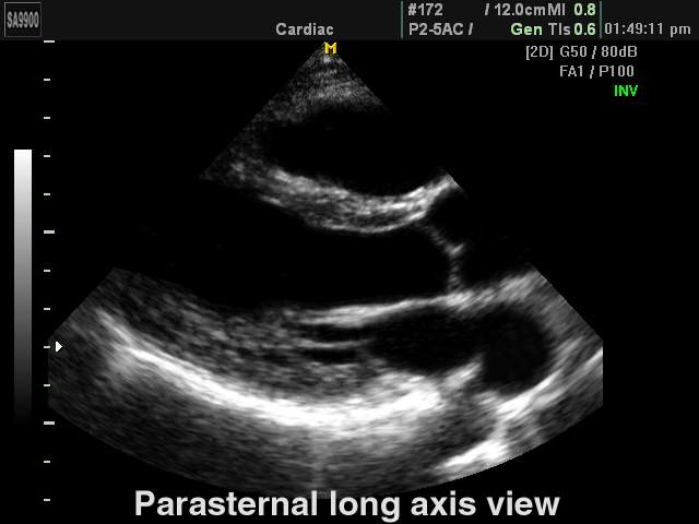
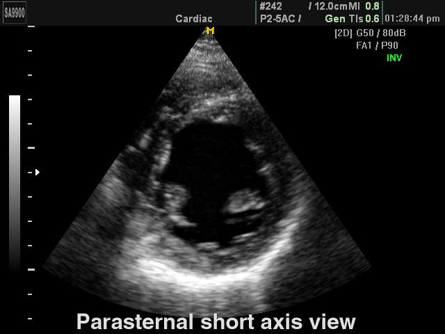
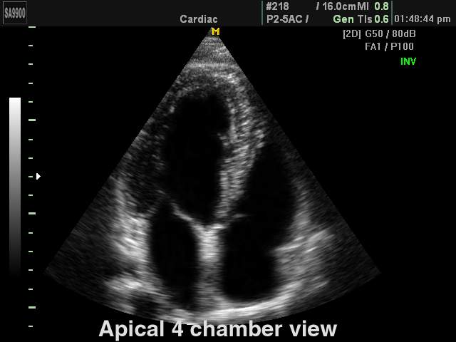
- Пакет кардиологических исследований:
- в 2D-режиме рассчитываются значения таких параметров, как объем по методу Симпсона (Simpson), объем по площади и длине, двумерные характериcтики (например, фракция выброса левого желудочка) и масса левого желудочка;
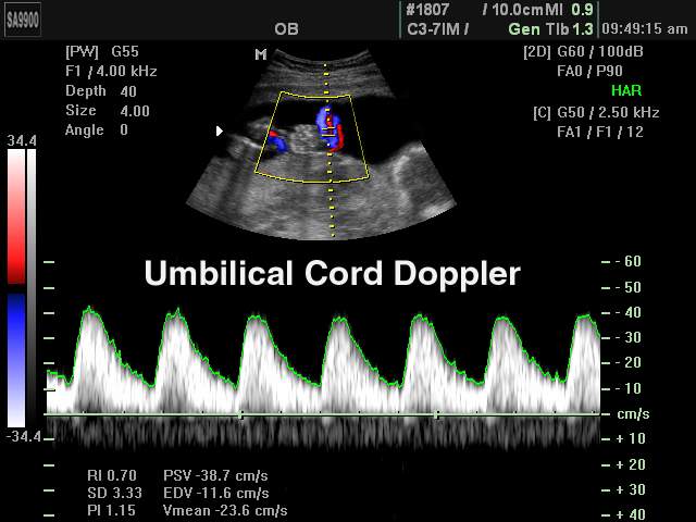
- в M-режиме вычисляются значения параметров для левого желудочка, аорты и левого предсердия, митрального клапана, а также частота сердечных сокращений. - Пакет расчетов параметров сосудов: вычисления объемного кровотока, процента стеноза, индекса сопротивления (RI), пульсационного индекса (PI) и др.
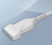
Датчики для УЗ сканера SonoAce-9900
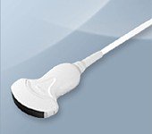
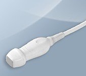
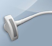
Конвексные датчики
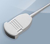
Конвексный датчик C 2-5EL/40/85
Акушерские исследования (плод, сердце плода), гинекология (матка, яичники), абдоминальные исследования (печень, желчный пузырь, поджелудочная железа, селезенка, глубокие сосуды), почки.
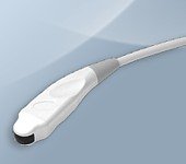
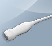
Конвексный датчик C 2-5ET/40/76
Акушерские исследования (плод, сердце плода), гинекология (матка, яичники), абдоминальные исследования (печень, желчный пузырь, поджелудочная железа, селезенка, глубокие сосуды), почки.
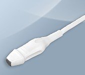
Конвексный датчик C 3-7ED/50/77
Акушерские исследования (плод, сердце плода), гинекология (матка, яичники), абдоминальные исследования (печень, желчный пузырь, поджелудочная железа, селезенка, глубокие сосуды), почки.
Конвексный датчик C 4-9ED/10/150 (микроконвексный)
Неонатология и педиатрия: абдоминальные исследования, почки, сердце, глубокие сосуды, мозг.
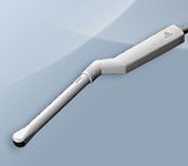
Конвексный датчик EC 4-9ED/10/150 (вагинальный)
Акушерские исследования (ранние сроки), гинекология (матка, яичники), урология (предстательная железа), исследования прямой кишки.
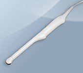
Конвексный датчик EC 4-9ES/10/150 (ректо-вагинальный)
Акушерские исследования (ранние сроки), гинекология (матка, яичники), урология (предстательная железа), исследования прямой кишки.
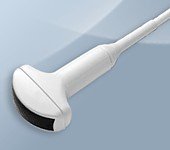
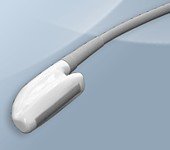
Фазированные датчики
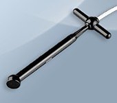
Фазированный датчик P 2-5AC/15
Кардиология и транскраниальные исследования у взрослых.
Фазированный датчик P 2-5AC/19
Кардиология и транскраниальные исследования у взрослых.
Фазированный датчик P 3-7AC/10
Кардиология и транскраниальные исследования у детей.
Линейные датчики
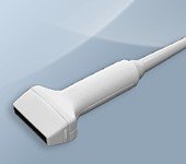
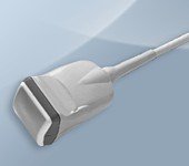
Линейный датчик L 5-12IM/40
Поверхностные структуры (щитовидная железа, молочная железа, лимфоузлы), мускулоскелетные исследования (суставы, мышцы, подкожные структуры), периферические сосуды.
Линейный датчик L 5-9ER/50
Поверхностные структуры (щитовидная железа, молочная железа, лимфоузлы), мускулоскелетные исследования (суставы, мышцы, подкожные структуры), периферические сосуды.
Интраоперационные датчики
Интраоперационный датчик CL 4-8/40/65
Абоминальные исследования во время операции.
Интраоперационный датчик LI 5-9EV/40
Абоминальные исследования во время операции.
Объемные датчики
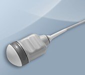
Объемный датчик 3D 3-5EK/40/69
Трехмерные абдоминальные исследования, акушерство и гинекология.

Объемный датчик 3D 4-8ET/40/84
Трехмерные абдоминальные исследования, акушерство (трехмерное УЗИ плода) и гинекология.

Объемный датчик 3D 5-8EK/10/128 (ректо-вагинальный)
Трехмерные исследования в акушерстве (ранние сроки), гинекологии (матка, яичники), урологии (предстательная железа), исследования прямой кишки.

Объемный датчик VAW 4-7/40/69
Трехмерные абдоминальные исследования, акушерство (трехмерное УЗИ плода) и гинекология.
Объемный датчик VDW 5-8/10/128 (ректо-вагинальный)
Трехмерные исследования в акушерстве (ранние сроки), гинекологии (матка, яичники), урологии (предстательная железа), исследования прямой кишки.
Объемный датчик VNW 6-12/40
Трехмерные исследования поверхностных структур (щитовидной железы, молочной железы, лимфоузлов), мускулоскелетные исследования (суставы, мышцы, подкожные структуры), периферические сосуды.
Допплеровские датчики
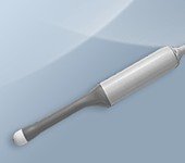
Допплеровский датчик CW 2.0 (слепой допплер)
Транскраниальные исследования, сосуды.
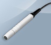
Допплеровский датчик CW 4.0 (слепой допплер)
Транскраниальные исследования, сосуды.
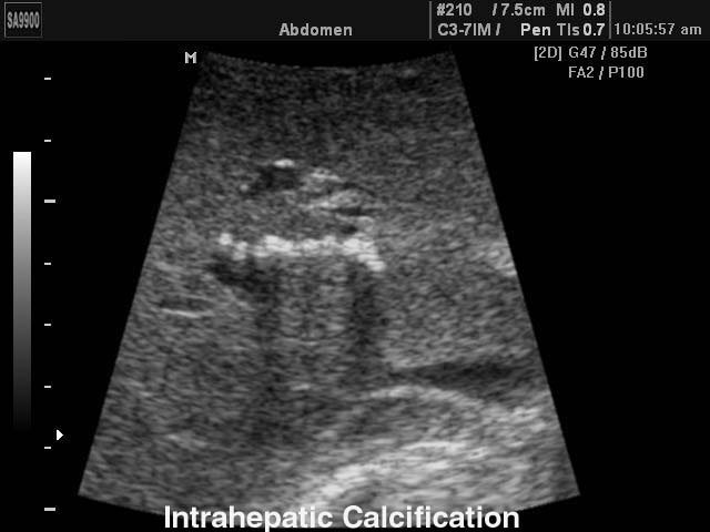
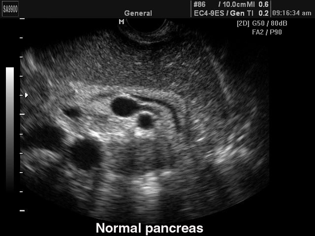
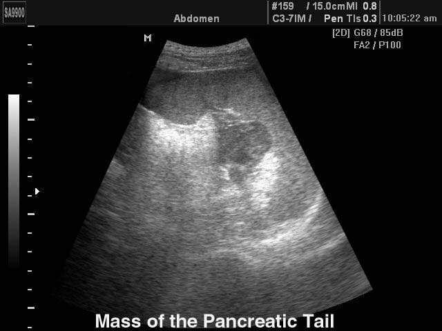
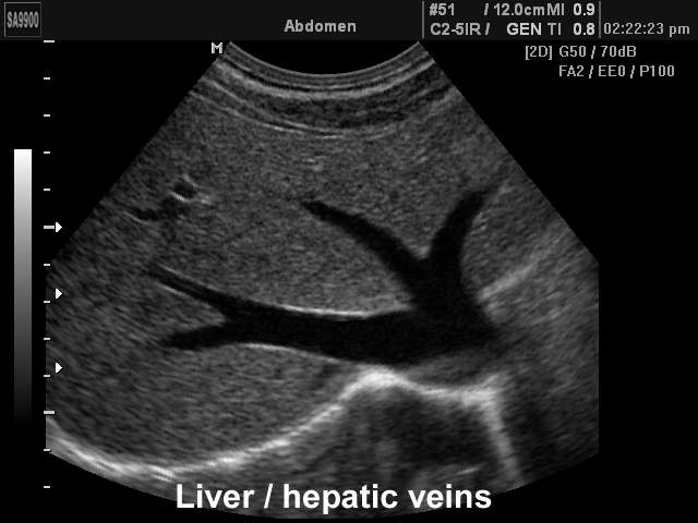
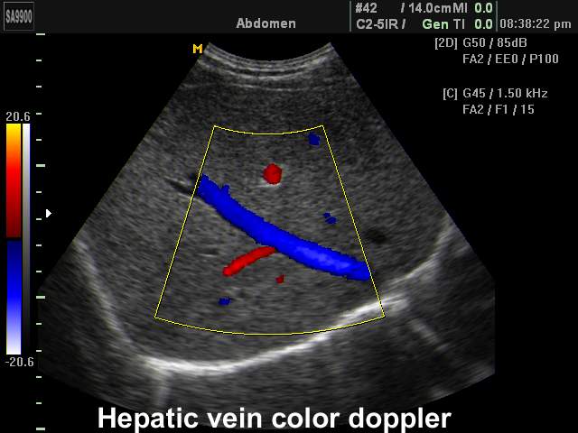
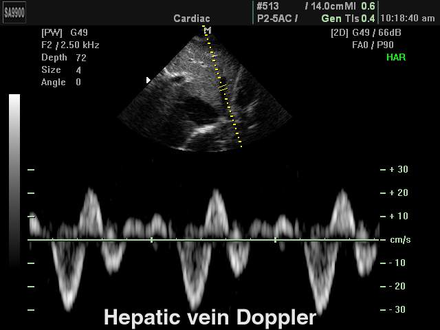
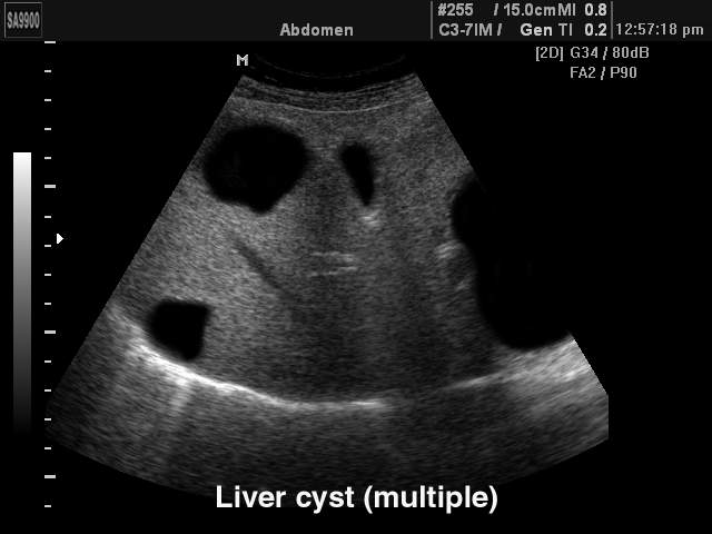
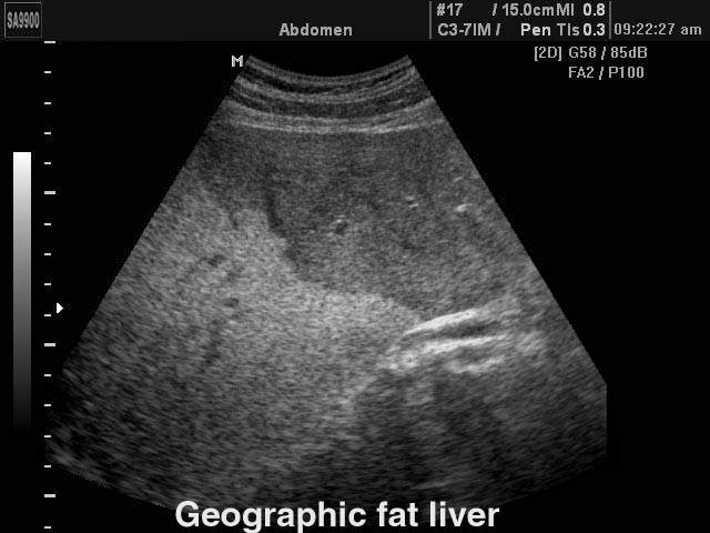
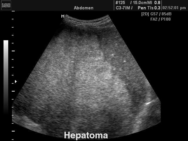
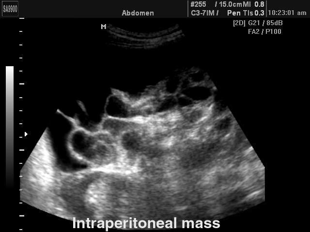
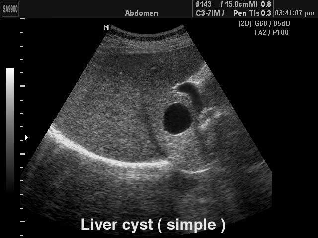
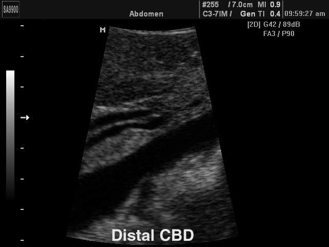
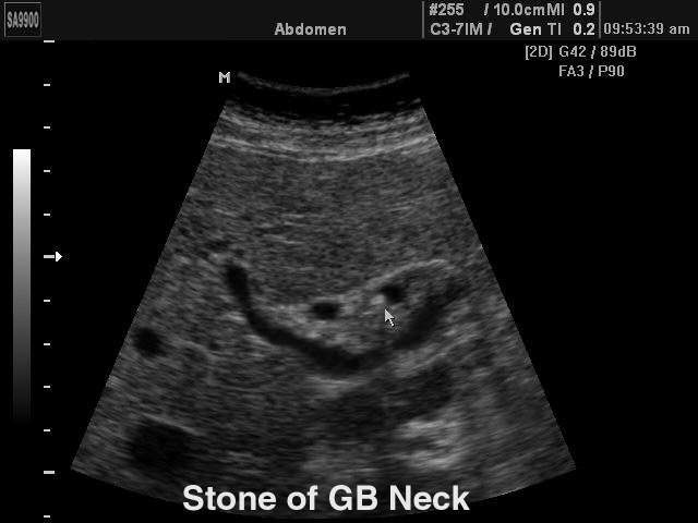
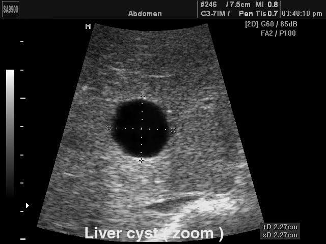
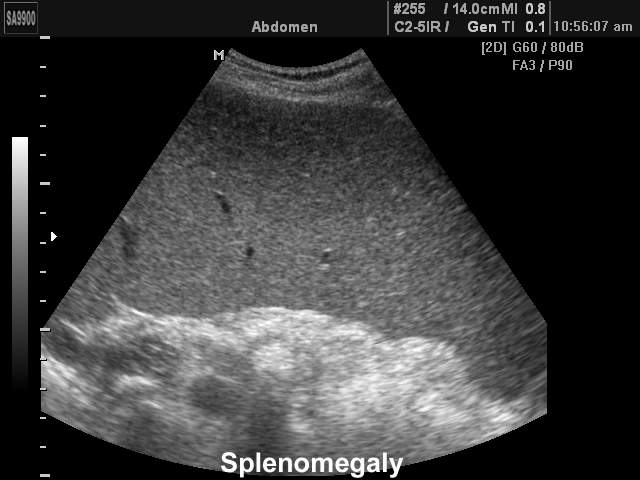
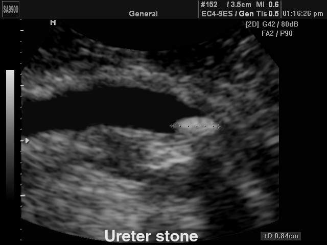
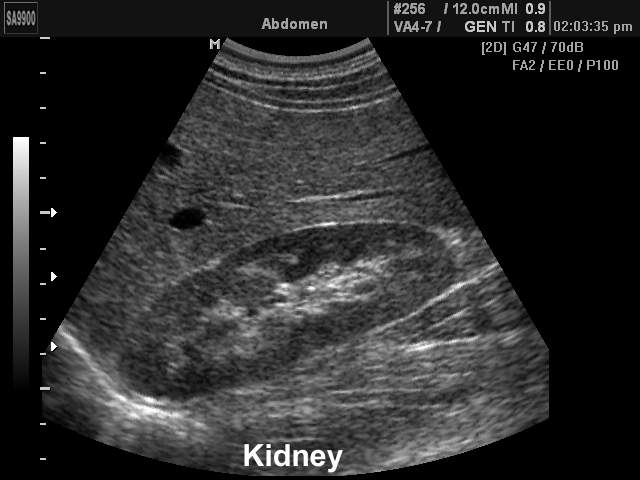
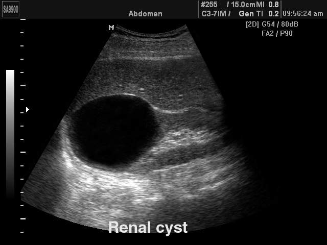
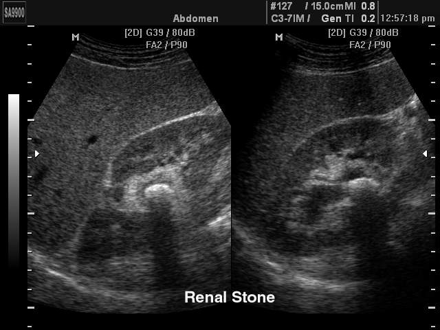
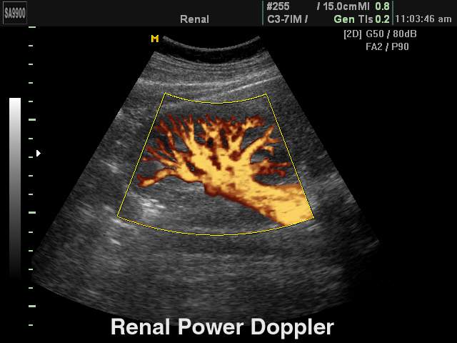
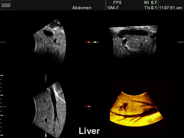
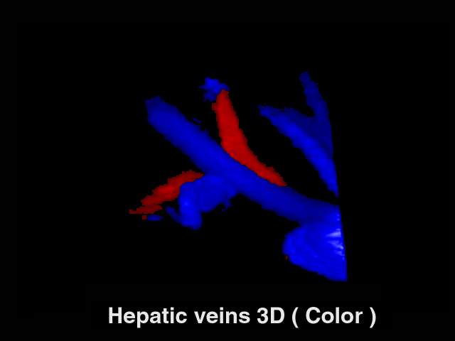
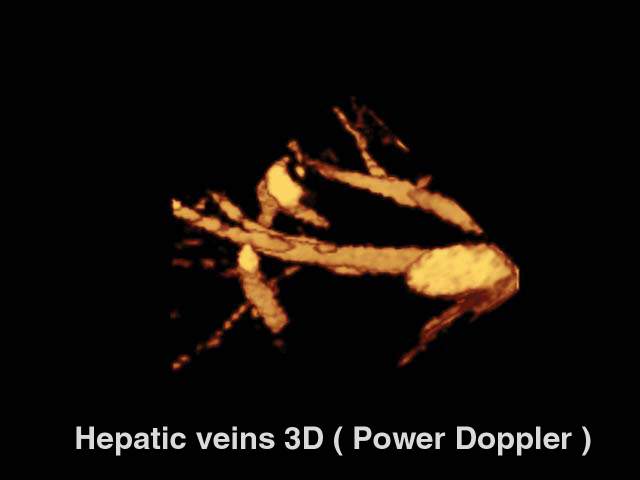
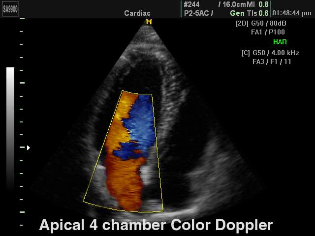
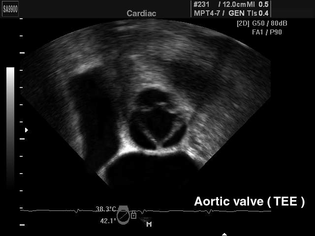
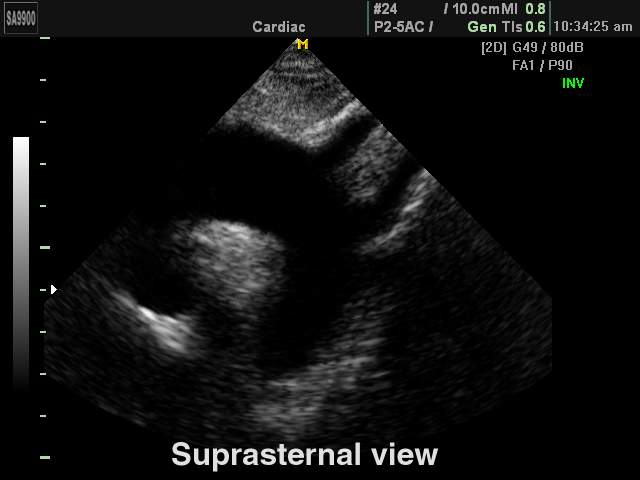
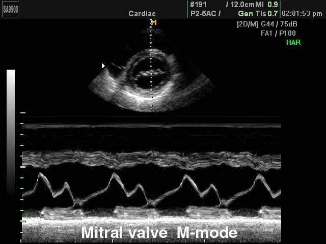
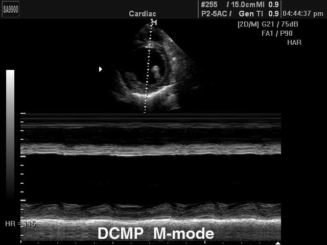
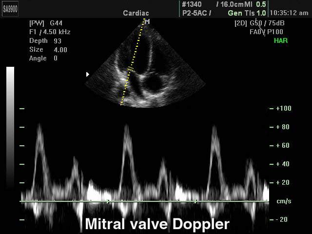
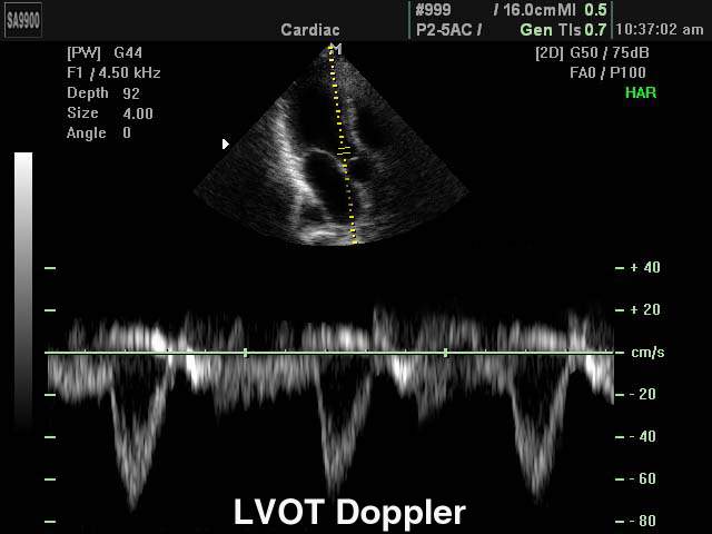
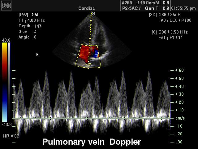
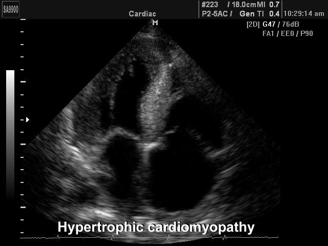
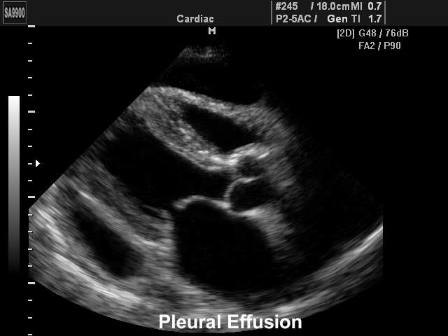
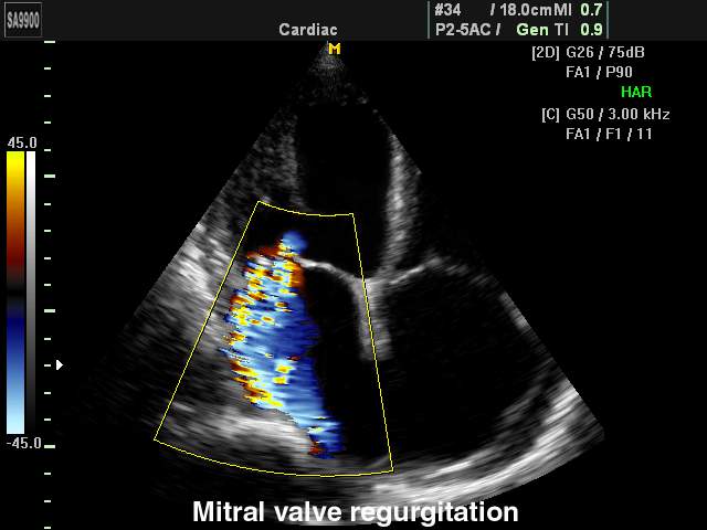
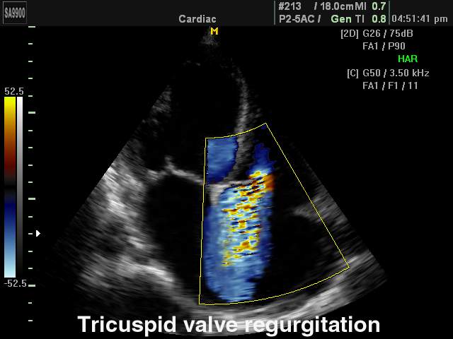
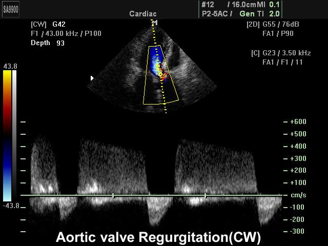
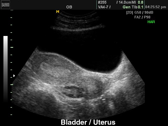
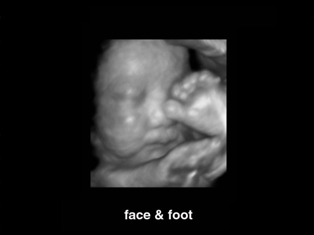
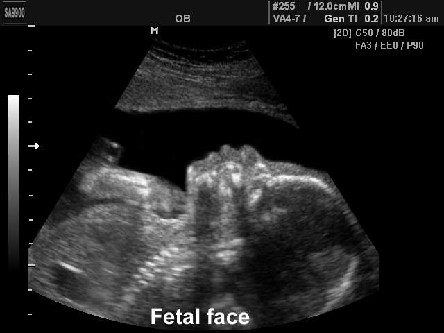
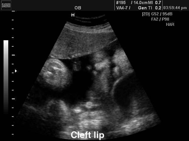
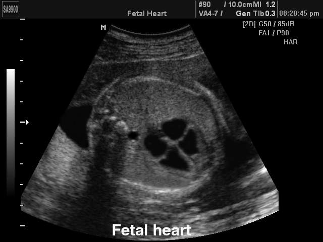
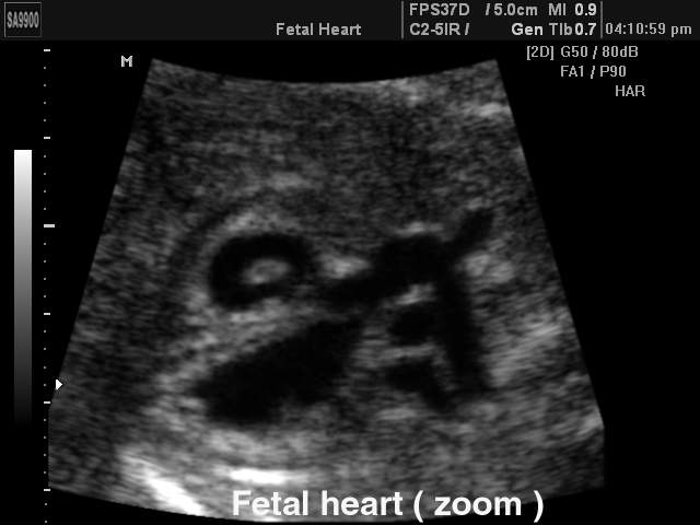
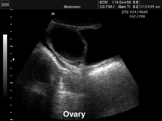
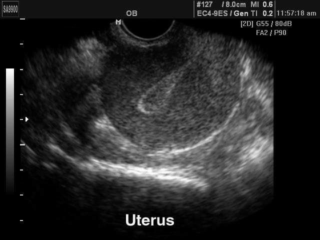
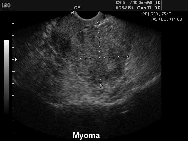
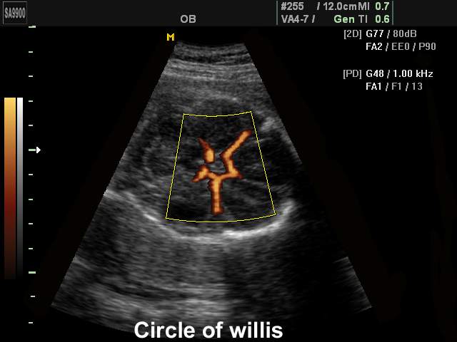
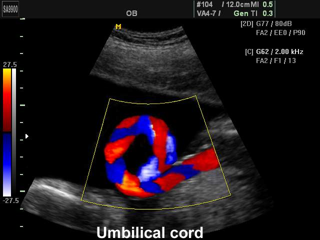
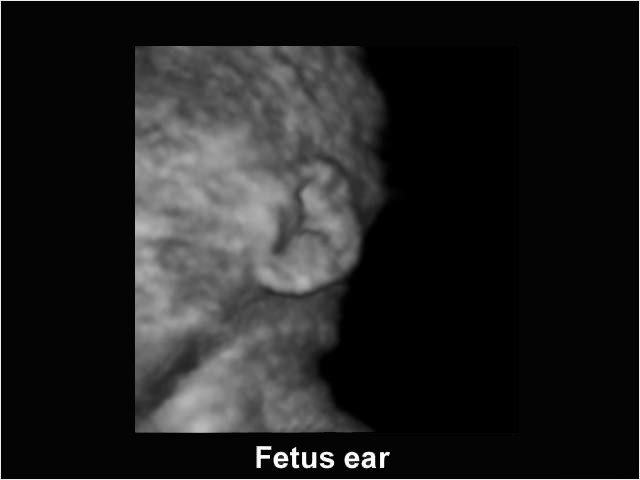
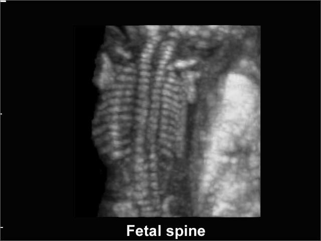
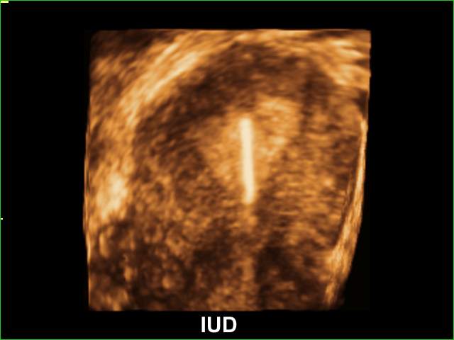
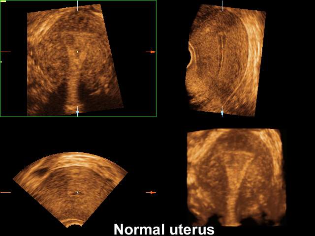
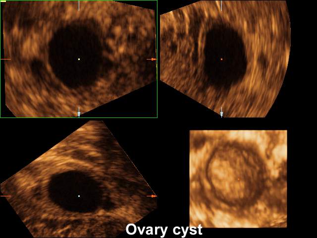
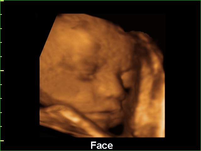
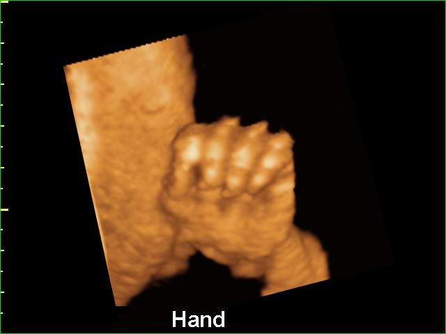
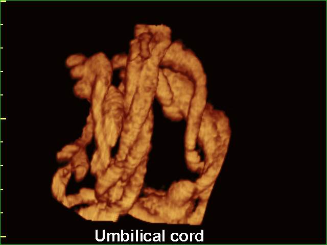
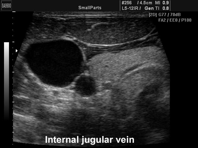
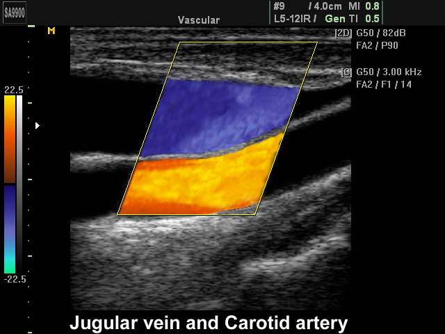
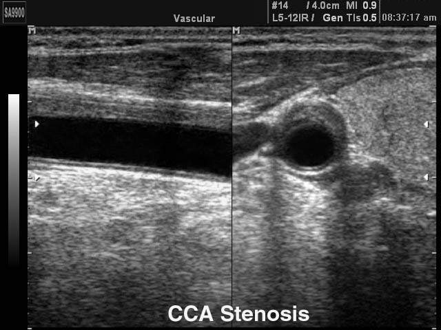
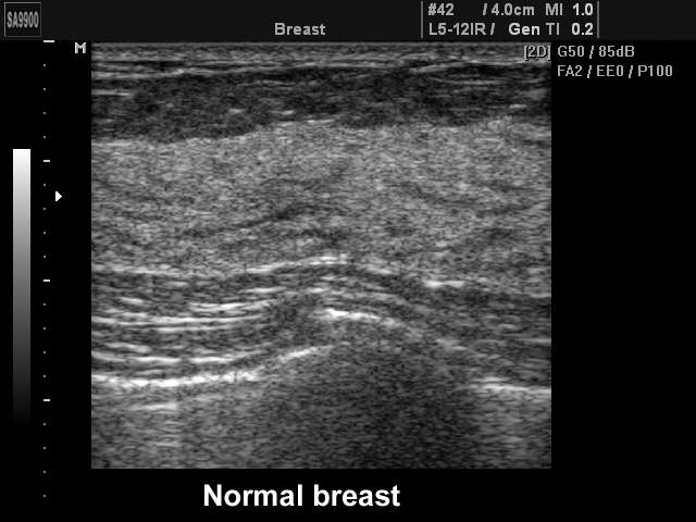
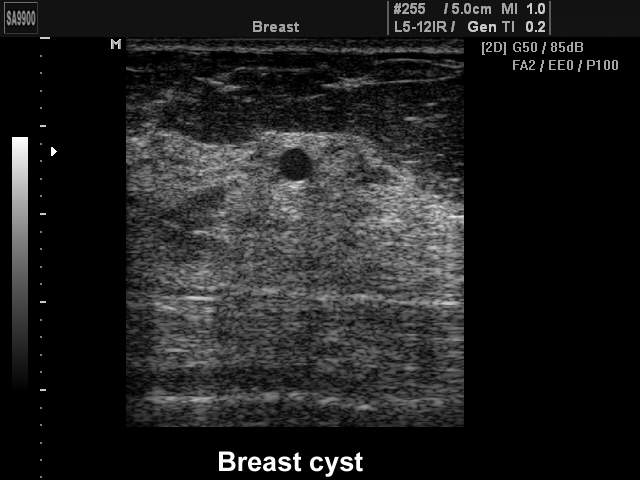
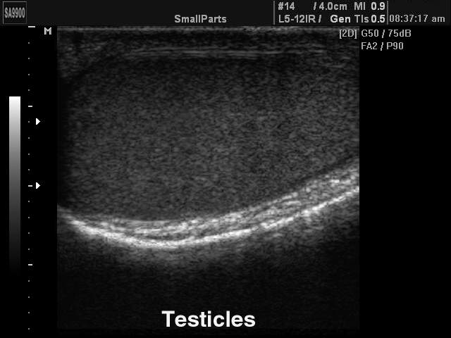
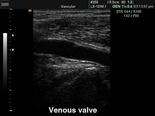
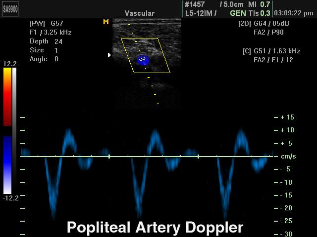
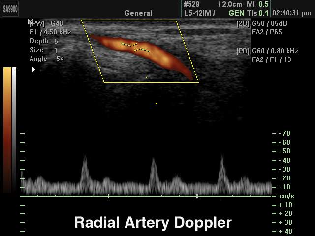
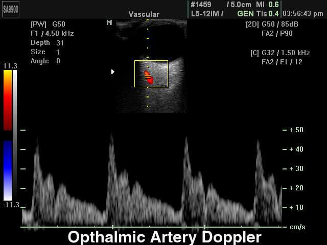
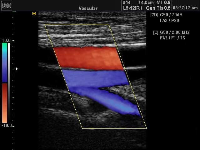
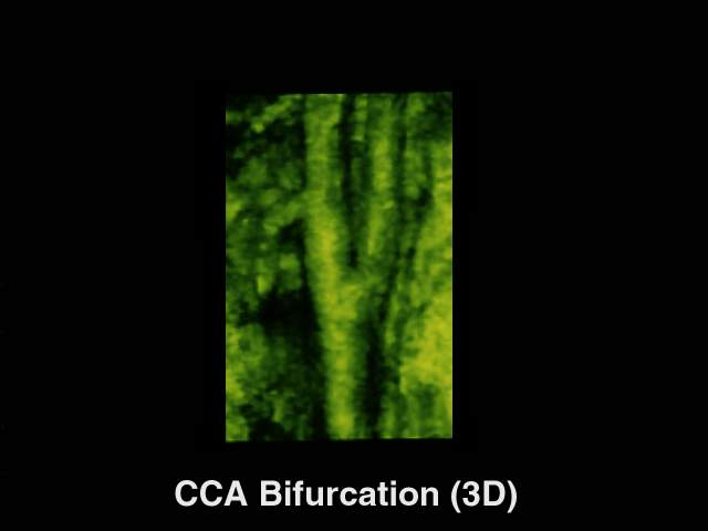
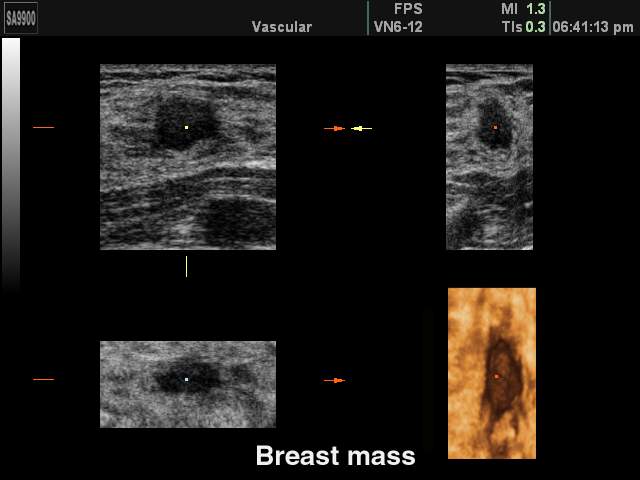
Примеры УЗИ на SonoAce-9900 (эхограммы)
Внимание! Копирование или частичная перепечатка материалов проспекта сканера "SonoAce 9900" на русском языке и размещение в интернет без согласия владельца (ЗАО "Медиэйс") запрещена, запрос на копирование материала можно отправить здесь.