
Atlas of ultrasound images - vessels (pg. 3)
In the section "Sonography of vessels" of atlas you can see echograms of examinations of organs and vessels with the usage of color and power mapping, continuous, pulsed and tissue dopplers.

SonoAce-X6. Common carotid artery - bifucation, B-mode.
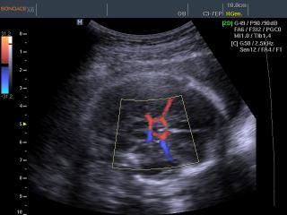
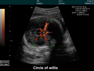
SonoAce-X6. Circle of Willis, color doppler.
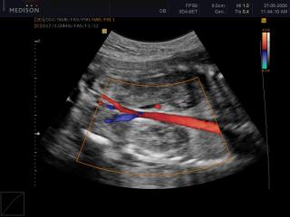
Accuvix-V20. Aorta, color doppler.
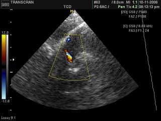
SonoAce-8000. Medial cerebral artery, color doppler.
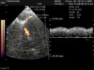
SonoAce-8000. Medial cerebral artery, PD & PW.
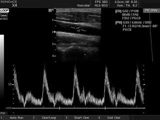
SonoAce-X4. Common carotid artery, PW doppler.
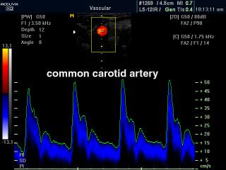
Accuvix-XQ. Common carotid artery, CFM & PW.
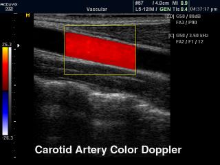
Accuvix-XQ. Common carotid artery, color doppler.
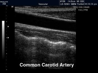
Accuvix-XQ. Common carotid artery, B-mode.
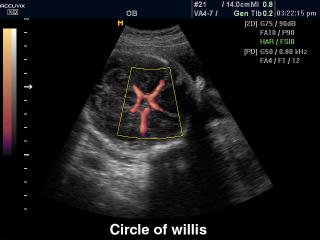
Accuvix-XQ. Circle of Willis, power doppler.
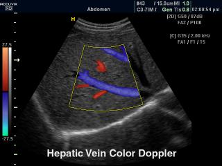
Accuvix-XQ. Hepatic vein, color doppler.
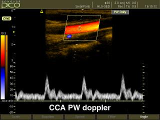
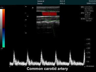
SonoAce-Pico. Common carotid artery, CFM & PW.
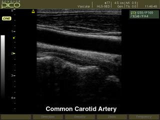
SonoAce-Pico. Common carotid artery, B-mode.

SonoAce-Pico. Circle of Willis, power doppler.
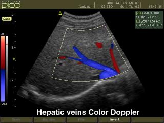
SonoAce-Pico. Hepatic vein, color doppler.
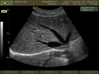
SonoAce-Pico. Hepatic vein, B-mode.
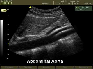
SonoAce-Pico. Abdominal aorta, B-mode.
SonoAce-6000 CMT. Common carotid artery, CFM & PW.
SonoAce-6000 CMT. Circle of Willis, power doppler.
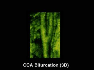
SonoAce-9900. Common carotid artery bifurcation, 3D.
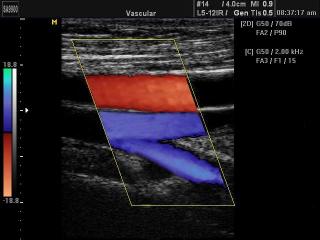
SonoAce-9900. Superficial femoral artery, color doppler.
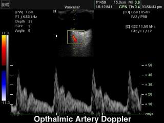
SonoAce-9900. Opthalmic artery, CFM & PW.
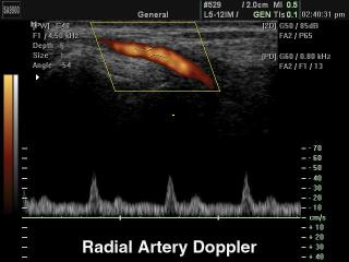
SonoAce-9900. Radial artery, PD & PW.
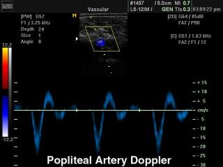
SonoAce-9900. Popliteal artery and vein, CFM & PW.
The description of ultrasound examinations in russian (эхография) and in english are situated in the database. The description of the diagnostic in the english language is closer to latin and doctors are likely to prefer this variant.