
Atlas of ultrasound images - vessels (pg. 4)
In the section "Sonography of vessels" of atlas you can see echograms of examinations of organs and vessels with the usage of color and power mapping, continuous, pulsed and tissue dopplers.
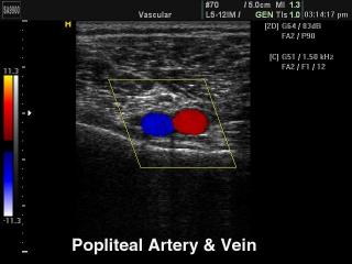
SonoAce-9900. Popliteal аrtery and vein, color doppler.
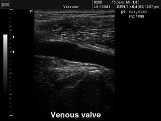
SonoAce-9900. Venous valve, B-mode.
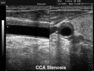
SonoAce-9900. Common carotid artery stennosis, B-mode.
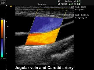
SonoAce-9900. Jugular vein and сarotid artery, color doppler.
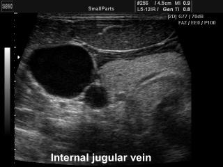
SonoAce-9900. Internal jugular vein, B-mode.
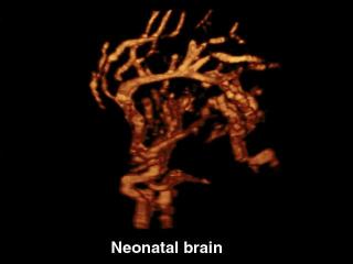
SonoAce-9900. Neonatal brain, power doppler & 3D.
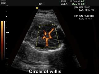
SonoAce-9900. Circle of Willis, power doppler.
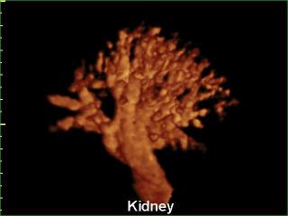
SonoAce-9900. Kidney vessels, power doppler, 3D.
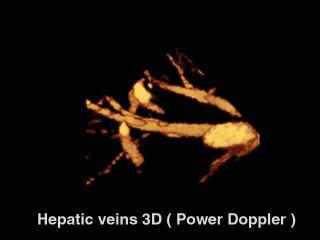
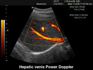
SonoAce-9900. Hepatic veins, power doppler, 3D.
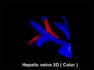
SonoAce-9900. Hepatic veins, color doppler, 3D.
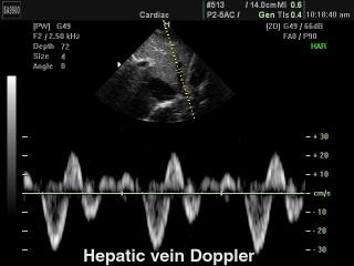
SonoAce-9900. Hepatic vein, PW.
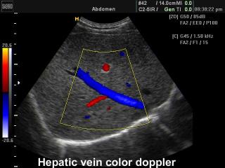
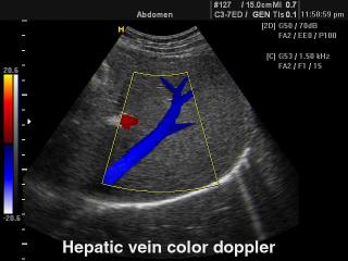
SonoAce-9900. Hepatic vein, color doppler.
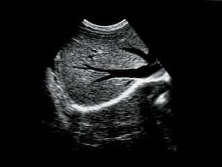
SonoAce-9900. Liver and hepatic veins, B-mode.

SonoAce-8000. Common carotid artery, CFM & PW.
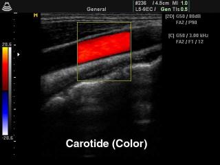
SonoAce-8000. Common carotid artery, color doppler.
SonoAce-8000. Common carotid artery, B-mode.
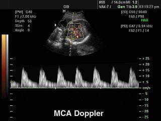
SonoAce-8000. Fetal middle cerebral artery, PD & PW.
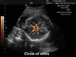
SonoAce-8000. Circle of Willis, power doppler.
SonoAce-8000. Hepatic vein, color doppler.
SonoAce-8000. Hepatic veins, power doppler.
SonoAce-600. Liver and hepatic veins, B-mode.
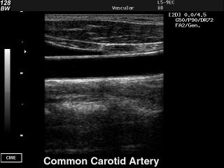
SonoAce-128BW. Common carotid artery, B-mode.
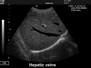
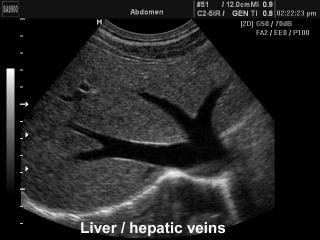
SonoAce-128BW. Hepatic veins, B-mode.
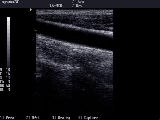
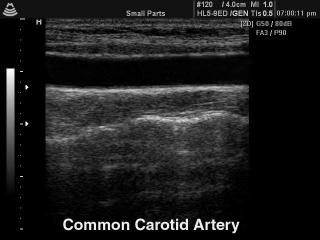
MySono-201. Common carotid artery, B-mode.
The description of ultrasound examinations in russian (эхография) and in english are situated in the database. The description of the diagnostic in the english language is closer to latin and doctors are likely to prefer this variant.