
Atlas of ultrasound images - vessels (pg. 5)
In the section "Sonography of vessels" of atlas you can see echograms of examinations of organs and vessels with the usage of color and power mapping, continuous, pulsed and tissue dopplers.
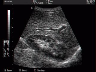
MySono-201. Umbilical artery, B-mode.
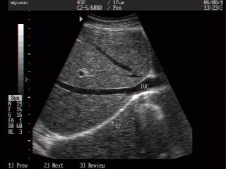
MySono-201. Inferior vena cava, B-mode.
The description of ultrasound examinations in russian (эхография) and in english are situated in the database. The description of the diagnostic in the english language is closer to latin and doctors are likely to prefer this variant.