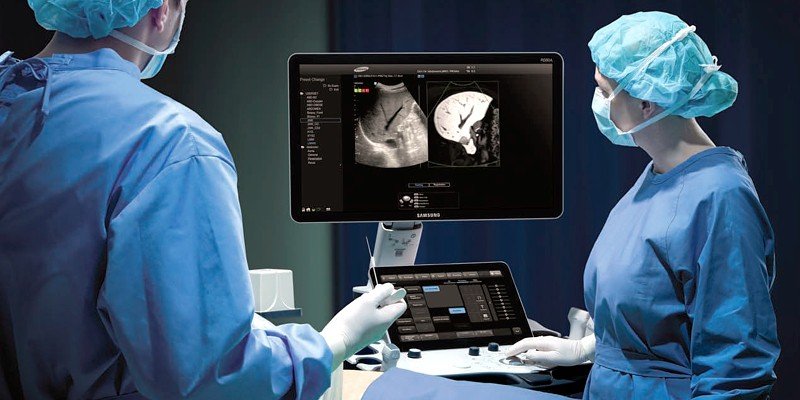
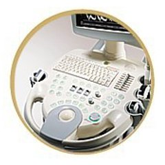
УЗИ сканер SonoAce-X4 (Medison, снят с производства)

SonoAce X4 - многофункциональный ультразвуковой сканер компании Medison с импульсным ч/б допплером (премиум класса). Аппарат позволяет проводить обследование большого потока пациентов - рекомендуется для применения в медицинских центрах, женских консультациях, больницах и поликлиниках.
Базовая комплектация: сканер SonoAce X4 (монитор 12,5"; кардиопакет для исследования сердца у взрослых и у плода; встроенные модули: импульсный допплер (PW), 2-я гармоника, FreeHand 3D, SonoView Lite; встроенный CD-RW дисковод; встроенная клавиатура с трекболом), флакон геля 250 мл и руководство оператора.
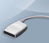
Опции для сканера SonoAce X4: разветвитель датчиков; устройство хранения информации - USB флеш-карта; система DICOM.
Области применения: акушерство и гинекология, абдоминальные исследования и маммология, урология и эхокардиография, поверхностно расположенные органы и мускуло-скелетные исследования, а также педиатрия..
Основные характеристики УЗ сканера SonoAce X4
- Стационарный ультразвуковой аппарат.
- Монитор - 12,5" (31 см).
- Режимы сканирования: B, 2B, B+M, B+2M, B+D.
- Увеличение в реальном масштабе времени, кинопамять.
- Разъемы для одновременного подключения 2-х датчиков.
- Особенности сканирования:
- тканевая гармоника (регистрация 2-й гармоники эхосигнала, в том числе с помощью инверсной технологии);
- автоматический анализ допплеровских кривых;
- PW - импульсный допплер;
- HPRF - высокочастотный импульсный допплер;
- глубина сканирования до 30 см. - Система FreeHand 3D - модуль трехмерной реконструкции, восстановление объемной структуры поверхностей тканей (функции увеличения, вращения и т.д.).
- Система SonoView Lite - система архивации и дальнейшего просмотра изображений. Имеется возможность проведения измерений в архиве. При наличии соответствующих приводов возможно копирование изображений на гибкие дискеты, компакт-диски, магнитооптику.
- Система DICOM (опция) - возможность сетевой интеграции с PACS-системами поддерживающими стандарт DICOM (например, для архивации или печати ультразвуковых эхограмм на оборудовании других производителей медтехники).
Инновационные технологии
- Multi-beam (мульти-луч) - технология цифрового формирования луча (устранение многократного отражения, нелинейного ослабления и неточного времени задержки в отличие от аналоговых систем).
- OTI (Optimum Tissue Imaging) - технология получения оптимального изображения тканей, благодаря коррекции скорости. Функция при помощи которой пользователь может выбрать оптимальную скорость для каждой области исследований, тем самым получая одновременно высокое качество изображений различных видов тканей, таких как жир, мышцы или паренхима печени.
- THI (Tissue Harmonic Imaging), тканевая или 2-я гармоника - повышает качество изображения линейное и контрастное разрешение у трудно визуализируемых пациентов. Данная технология предполагает использование широкополосных датчиков и приемного тракта повышенной чувствительности. Дает преимущество при исследовании пациентов с повышенным весом.
- OHI (Optimized Harmonic Imaging) - объединяет две предыдущие технологии и предназначена для особо трудных для визуализации случаев.
- FINE (Filtered Image for Noise reduction & Edge enhancement) - программа фильтрации ультразвукового изображения. Обеспечивает лучшую контрастность контуров и уменьшает уровень шумов.
- Quick Scan - ускоренный режим (нажатием одной кнопки) настройки изображения исследуемого органа в B-режиме и D-режиме (настройка оптимальных параметров и фильтров за счет автоматического распознавания исследуемого органа по интеллектуальной базе данных человеческих органов).
- FSI (Full Spectrum Imaging) - технология, которая объединяет ультразвуковую информацию от акустических полос разной частоты, что резко снижает количество артефактов и формирует превосходное изображение с плотной контрастностью и значительно лучшей степенью проникновения. Данная технология использовалась ранее только в аппаратах премиум класса (Accuvix), теперь она есть и в сканере SonoAce X4.
Пакеты диагностических программ, автоматические вычисления и количественные оценки
- Основные измерения: измерения расстояния, окружности, площади, объема; измерение тазобедренного сустава; измерение расстояния в M-режиме и др.
- Пакет гинекологических исследований: матка, левый и правый яичники, левая и правая почки, артерии левого и правого яичников, левый и правый фолликулы.
- Пакет акушерских исследований: биометрия плода, краниологическое исследование плода, исследование длинных костей плода, измерение индекса околоплодных вод (AFI) и др. Биометрия плода включает измерения теменно-копчиковой длинны (CRL), размера плодного пузыря (GS), бипариетальный размера головки плода (BPD), затылочно-лобного расстояния (OFD), длины окружности головы плода (НC), передне-заднего размера брюшной полости (APD), поперечного размера брюшной полости (TAD), окружности живота (AC), площади сечения тела (FTA), длины бедра (FL), поперечного (TTD) и передне-заднего (APTD) размеров тела плода. Краниологическое исследование плода включает измерения параметров мозжечка (CEREB), а также внешнего (OOD) и внутреннего (IOD) межглазных расстояний. Исследование длинных костей плода включает измерения длины плечевой кости (Humerus), локтевой кости (Ulna), большеберцовой кости (Tibia), лучевой кости (Rad), ключицы (Clav) и позвоночника (LV).
Кроме того, семь уравнений для оценки веса плода: Хедлок (Hadlock) 1-4, Хансман (Hansmann) и Мерц (Merz); ЧСС плода (Fetal HR); таблицы, определяемые пользователем. - Пакет урологических расчетов: разностный объем, объем предстательной железы, вычисление плотности простатспецифического антигена (PSA).
- Пакет кардиологических исследований:
В-режим: оценка сократительной способности миокарда модифицированным методом Симпсона (Simpson), методом "площадь-длина", оценка массы левого желудочка, соотношение камер сердца и т.д.
M-режим: оценка сократительной способности миокарда, размеров камер сердца и клапанов, измерение частота сердечных сокращений.
D-режим: оценка работы клапанного аппарата и исследование кровотока в сосудах (скорости, градиенты давления, индексы).
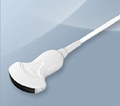
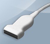
Датчики для сканера SonoAce X4
Конвексные датчики
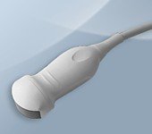
Конвексный датчик 2-4 МГц (микроконвексный)
Кардиология, абдоминальные исследования (печень, желчный пузырь, поджелудочная железа, селезенка, глубокие сосуды), почки.
Биопсийный набор: нет.
Конвексный датчик 2-5 МГц
Акушерские исследования (плод, сердце плода), гинекология (матка, яичники), абдоминальные исследования (печень, желчный пузырь, поджелудочная железа, селезенка, глубокие сосуды), почки.
Биопсийный набор: есть.
Конвексный датчик 3-7 МГц
Акушерские исследования (плод, сердце плода), гинекология (матка, яичники), абдоминальные исследования (печень, желчный пузырь, поджелудочная железа, селезенка, глубокие сосуды), почки.
Биопсийный набор: есть.

Конвексный датчик 4-9 МГц (ректо-вагинальный)
Акушерские исследования (ранние сроки), гинекология (матка, яичники), урология (предстательная железа), исследования прямой кишки.
Биопсийный набор: есть.
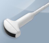
Линейные датчики
Линейный датчик 5-12 МГц
Поверхностные структуры (щитовидная железа, молочная железа, лимфоузлы), мускулоскелетные исследования (суставы, мышцы, подкожные структуры), периферические сосуды.
Биопсийный набор: есть.
Линейный датчик 5-9 МГц
Поверхностные структуры (щитовидная железа, молочная железа, лимфоузлы), мускулоскелетные исследования (суставы, мышцы, подкожные структуры), периферические сосуды.
Биопсийный набор: есть.
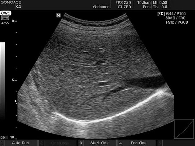
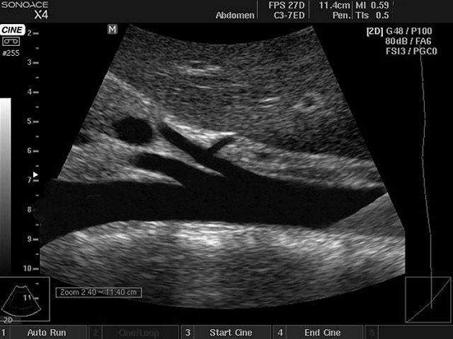
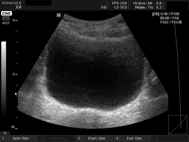
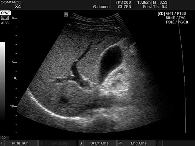
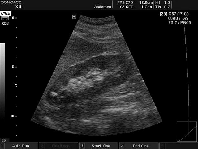
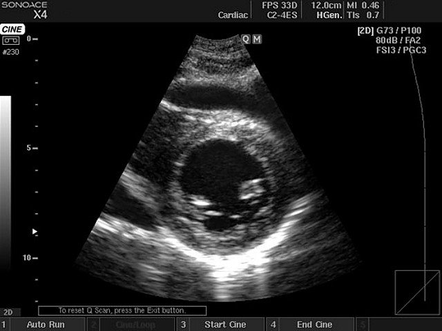
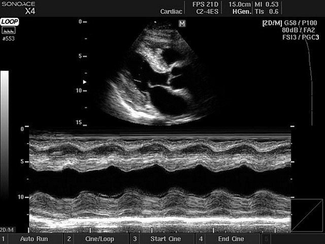
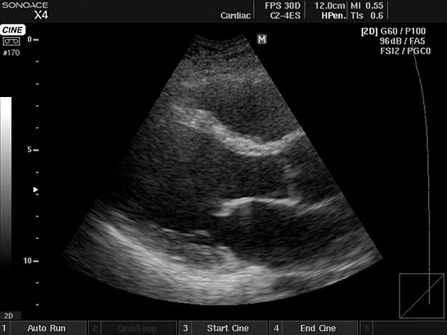
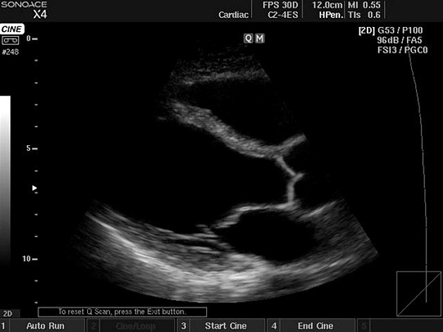
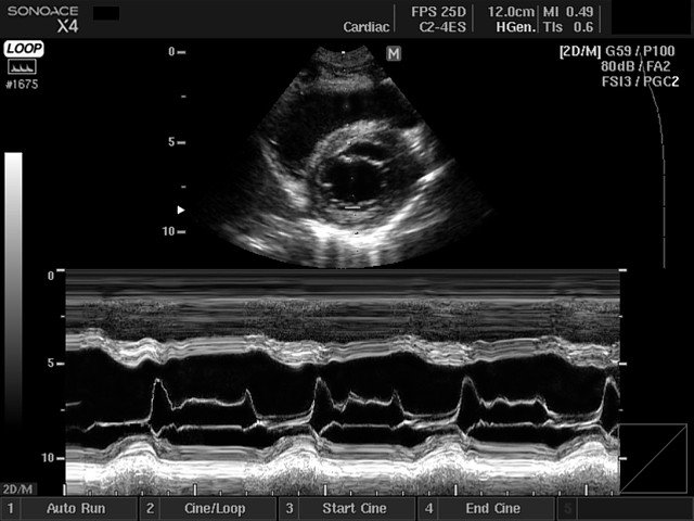
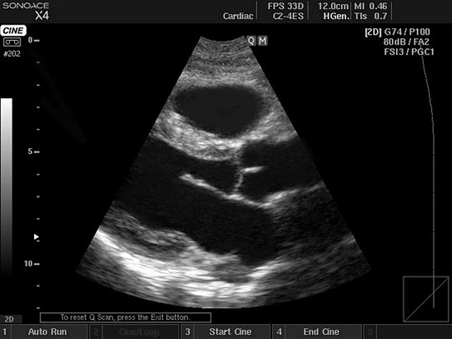
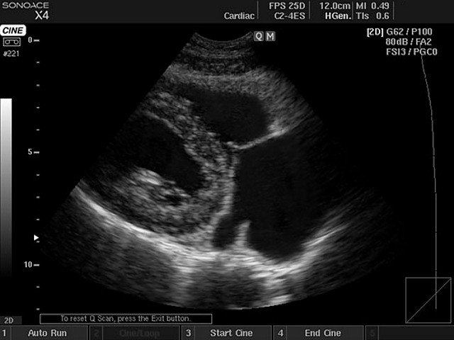
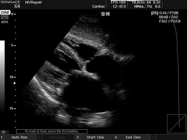
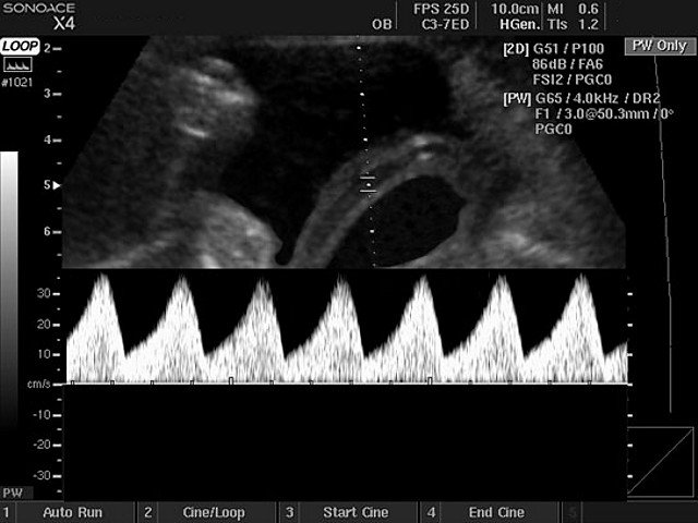
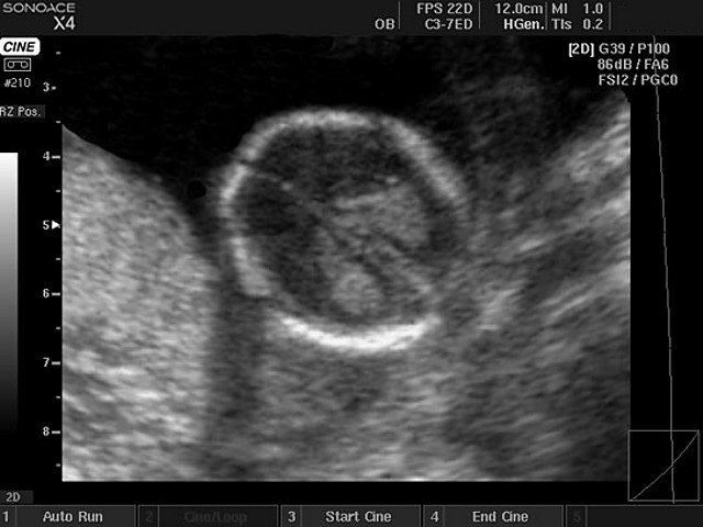
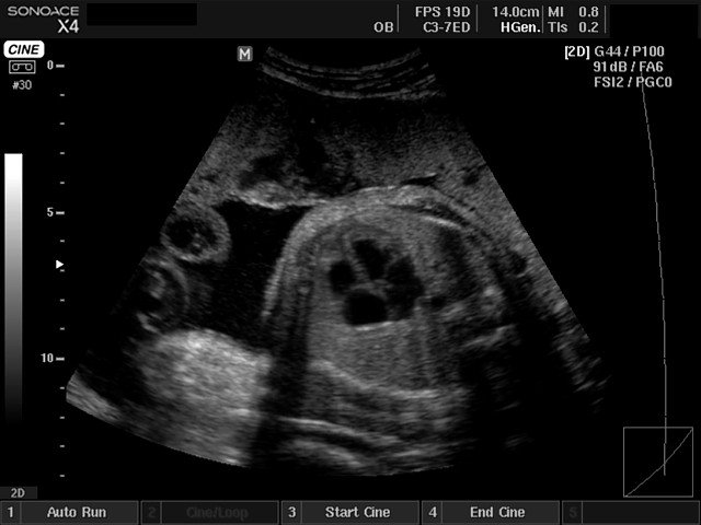
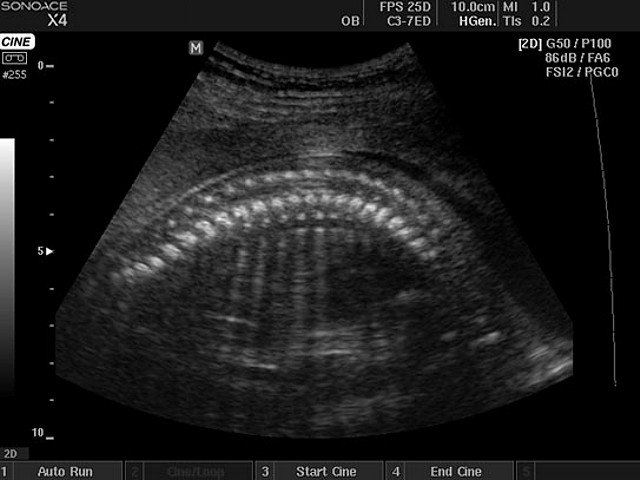
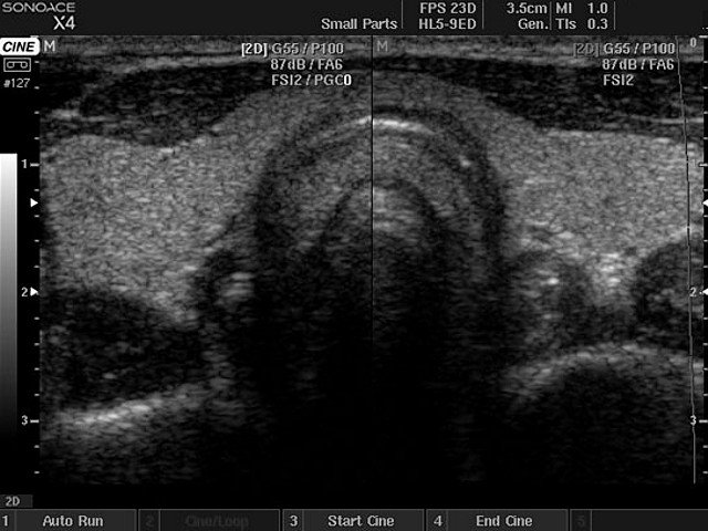
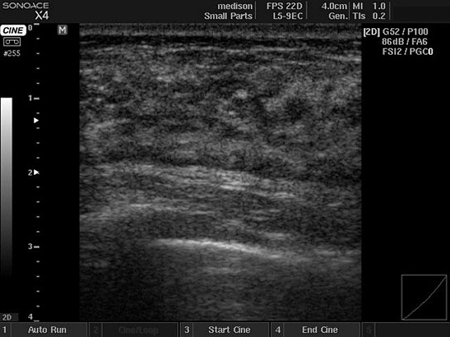
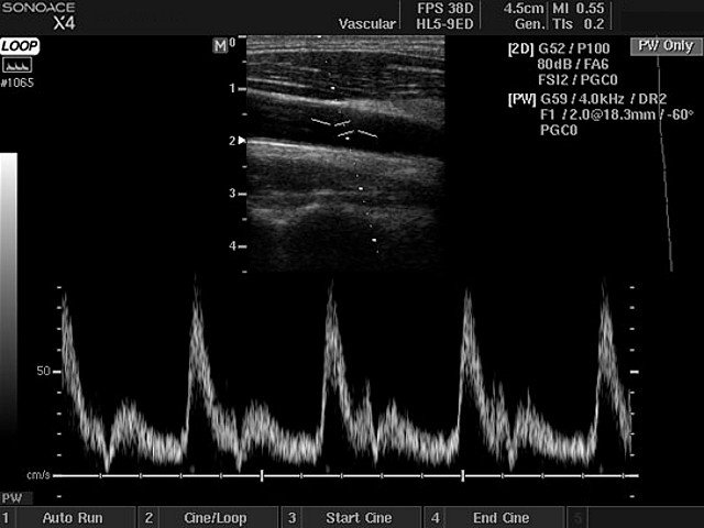
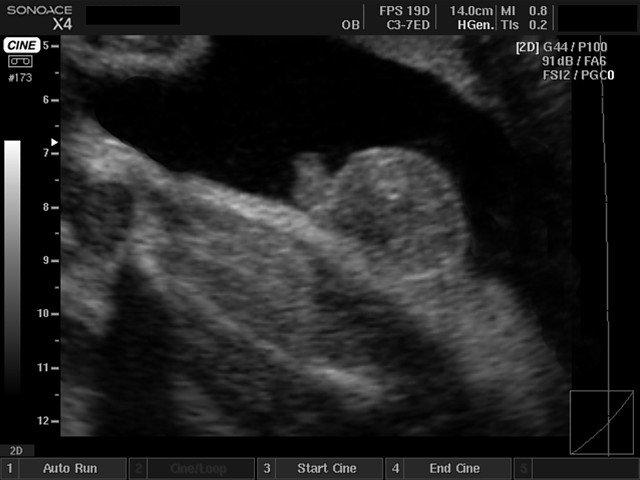
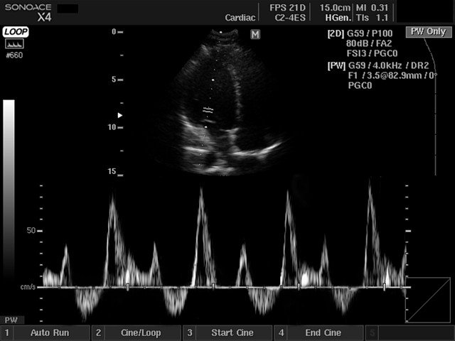
Примеры УЗИ на сканере SonoAce X4 (эхограммы)
Внимание! Копирование или частичная перепечатка материалов проспекта сканера "SonoAce X4" на русском языке и размещение в интернет без согласия владельца (ЗАО "Медиэйс") запрещена, запрос на копирование материала можно отправить здесь.