
Atlas of ultrasound images - echocardiography (pg. 4)
In this section of atlas the results of ultrasonic examinations in cardiology are represented. Here you can see images: aortic, mitral and tricuspid valves, left and right ventricles, echograms of cardiomyopathy, regurgitation of valves, etc.
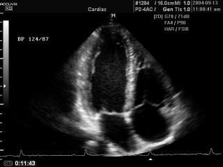
Accuvix-XQ. Heart (4 chamber view), B-mode (videо).
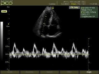
SonoAce-Pico. Mitral valve, CFM & PW.
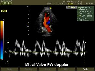
SonoAce-Pico. Mitral valve, PW.
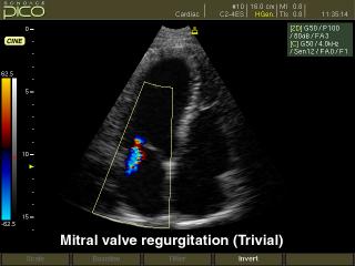
SonoAce-Pico. Mitral valve regurgitation, color doppler.
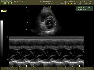
SonoAce-Pico. Mitral valve, M-mode.
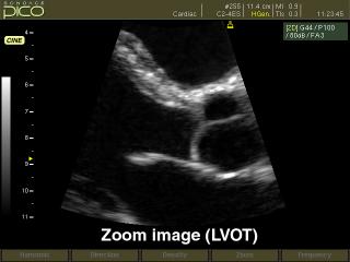
SonoAce-Pico. Left ventrical - outflow track, B-mode.
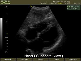
SonoAce-Pico. Heart (subcostal view), B-mode.
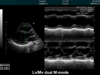
SonoAce-6000 CMT. Left ventricle and mitral valve, M-mode.
SonoAce-9900. Aortic valve regurgitation, CFM & CW.
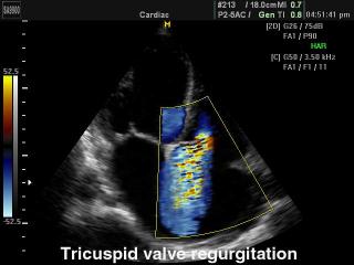
SonoAce-9900. Tricuspid valve regurgitation, color doppler.
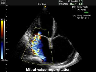
SonoAce-9900. Mitral valve regurgitation, color doppler.
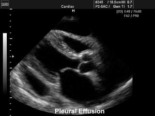
SonoAce-9900. Pleural effusion, B-mode.
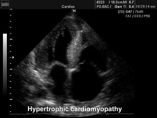
SonoAce-9900. Hypertrophic cardiomyopathy, B-mode.
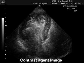
SonoAce-9900. Heart - contrast echography, B-mode.
SonoAce-9900. Heart, CFM & PW.
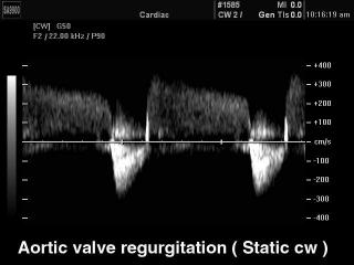
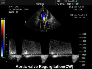
SonoAce-9900. Aortic valve regurgitation, CW.
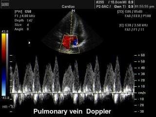
SonoAce-9900. Pulmonary vein, CFM & PW.
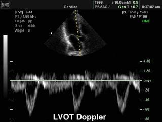
SonoAce-9900. Left ventricular - outflow tract, PW.
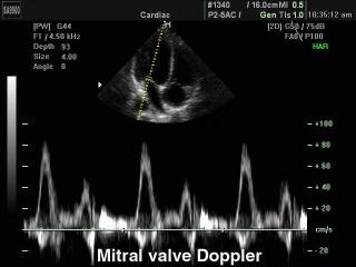
SonoAce-9900. Mitral valve, PW.
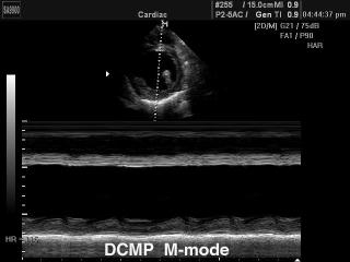
SonoAce-9900. Cardiomyopathy, M-mode.
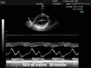
SonoAce-9900. Mitral valve, M-mode.
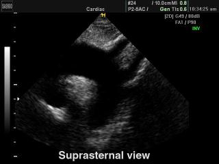
SonoAce-9900. Heart (suprasternal view), B-mode.
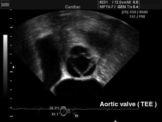
SonoAce-9900. Aortic valve (transesophageal echocardiography), B-mode.
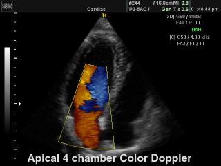
SonoAce-9900. Heart (4 chamber view), color doppler.
The description of ultrasound examinations in russian (эхография) and in english are situated in the database. The description of the diagnostic in the english language is closer to latin and doctors are likely to prefer this variant.