
Atlas of ultrasound images - echocardiography (pg. 3)
In this section of atlas the results of ultrasonic examinations in cardiology are represented. Here you can see images: aortic, mitral and tricuspid valves, left and right ventricles, echograms of cardiomyopathy, regurgitation of valves, etc.
SonoAce-8000. Additional hord of LV, B-mode.

SonoAce-8000. Aneurismus ascendus aorta, B-mode.
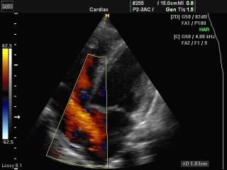
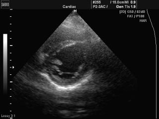
SonoAce-8000. Interatrium septum defect, color doppler.
SonoAce-8000. Mitral valve - myxomatous degeneration of cuspis, B-mode.
SonoAce-8000. Mitral valve prolapse, B-mode.
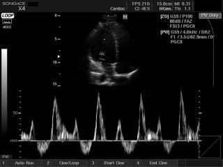
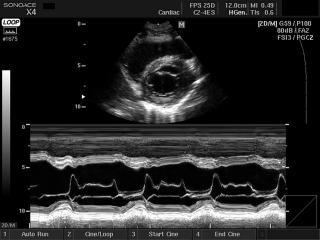
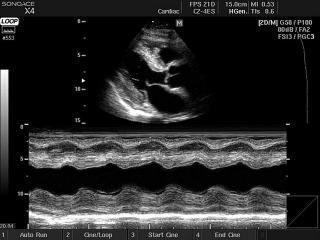
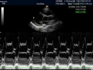
SonoAce-X4. Heart - transmitral inflow, PW doppler.
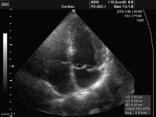
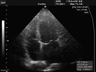
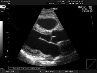
SonoAce-X4. Heart (subcostal view), B-mode.
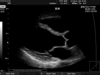
SonoAce-X4. Heart - right ventricular inflow, B-mode.
SonoAce-X4. Heart (long axis of LV), B-mode.
SonoAce-X4. Mitral valve, M-mode.
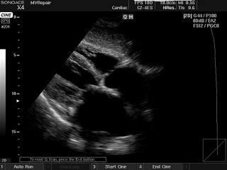
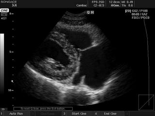
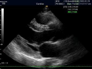
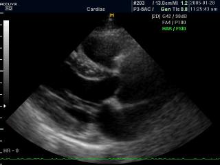
SonoAce-X4. Heart, B-mode.
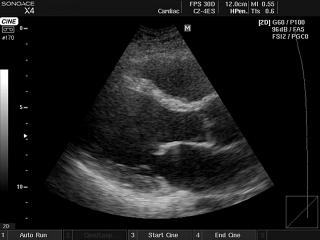
SonoAce-X4. Heart, B-mode.
SonoAce-X4. Heart - amyloidosis, M-mode.
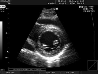
SonoAce-X4. Heart (short axis of LV), B-mode.
Accuvix-XQ. Mitral valve, anatomical M-mode.
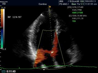
Accuvix-XQ. Heart, tissue doppler (videо).
Accuvix-XQ. Pulmanic artery, color doppler (videо).
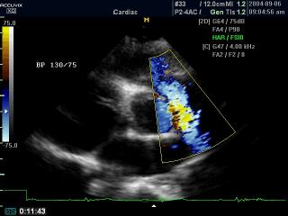
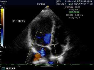
Accuvix-XQ. Mitral valve regurgitation, color doppler (videо).
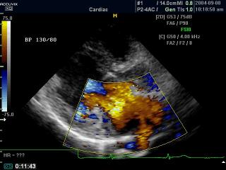
Accuvix-XQ. Heart, color dopler (videо).
Accuvix-XQ. Heart - pulse inversion harmonic, B-mode (videо).
Accuvix-XQ. Heart - tissue harmonic, B-mode (videо).
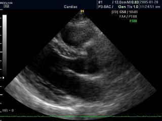
Accuvix-XQ. Heart - fundamental harmonic, B-mode (videо).
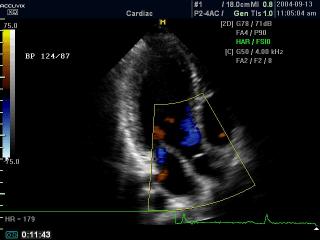
Accuvix-XQ. Mitral valve regurgitation, color doppler (videо).
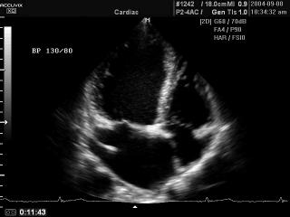
Accuvix-XQ. Mitral valve disease, B-mode (videо).
The description of ultrasound examinations in russian (эхография) and in english are situated in the database. The description of the diagnostic in the english language is closer to latin and doctors are likely to prefer this variant.