
Atlas of ultrasound images - echocardiography (pg. 2)
In this section of atlas the results of ultrasonic examinations in cardiology are represented. Here you can see images: aortic, mitral and tricuspid valves, left and right ventricles, echograms of cardiomyopathy, regurgitation of valves, etc.
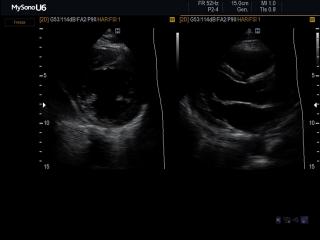
MySono-U6. Heart (short & long axis of the LV), B-mode.
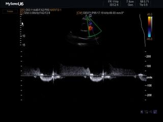
MySono-U6. Pulmonary regurgitation, CFM & CW.
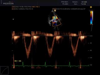
Accuvix-V10. Aortic valve, CFM & PW.
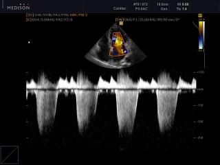
Accuvix-V10. Mitral valve - regurgitation, CFM & CW.
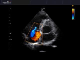
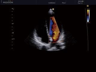
SonoAce-R7. Mitral valve - regurgitation, color doppler.
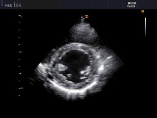
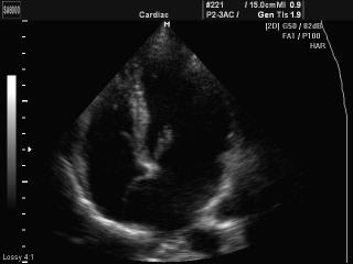
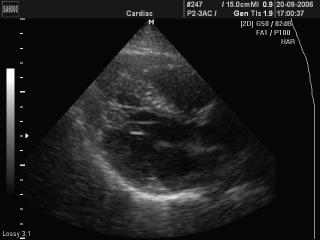
SonoAce-R7. Heart (short axis of LV), B-mode.
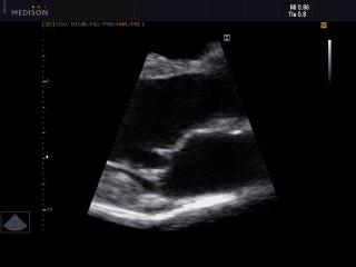
SonoAce-R7. Heart (long axis of LV), B-mode.
EKO7. Interventricular septum, TDI color doppler.
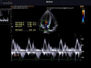
EKO7. Transmitral flow, CFM & PW.
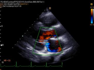
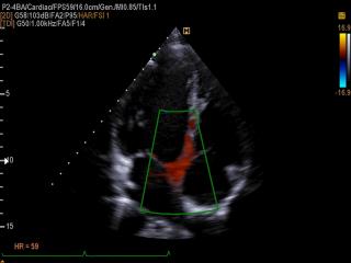
EKO7. Mitral regurgitation, color doppler.
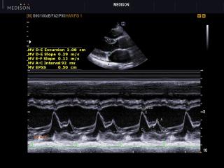
EKO7. Mitral valve, M-mode.
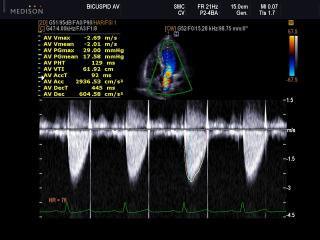
EKO7. Transaortic flow, CFM & CW.
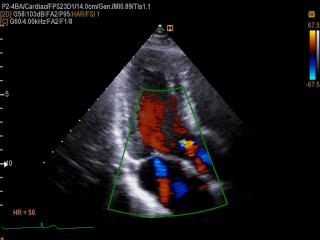
EKO7. Mitral regurgitation and aortic regurgitation, color doppler.
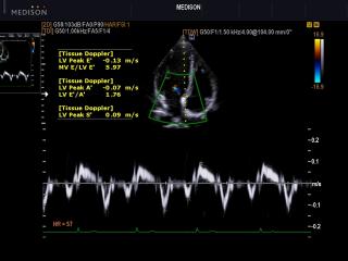
EKO7. Interventricular septum, TDI spectral doppler.
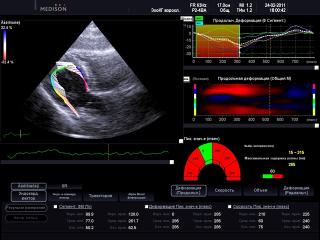
EKO7. Left ventricular longitudinal, 2D Strain and endocardial vectors.

EKO7. Left ventricular longitudinal, 2D Strain and acoustic markers.
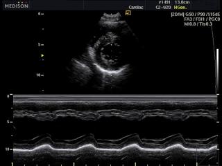
SonoAce-R3. Heart - left ventricle, M-mode.
MySono-U5. Heart (4 chamber view), color doppler.
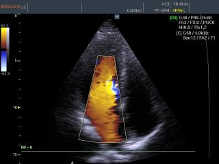
SonoAce-X6. Mitral valve, color doppler.
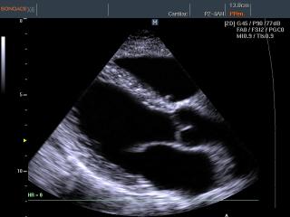
SonoAce-X6. Heart (long axis of LV), B-mode.
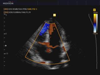
SonoAce-X8. Heart, tissue doppler.

SonoAce-8000. Aneurismus interatrium septum, B-mode.
SonoAce-8000. Aneurismus interatrium septum, B-mode.
SonoAce-8000. Additional muscles trabecula of LV, B-mode.
The description of ultrasound examinations in russian (эхография) and in english are situated in the database. The description of the diagnostic in the english language is closer to latin and doctors are likely to prefer this variant.