
Atlas of ultrasound images - echocardiography (pg. 5)
In this section of atlas the results of ultrasonic examinations in cardiology are represented. Here you can see images: aortic, mitral and tricuspid valves, left and right ventricles, echograms of cardiomyopathy, regurgitation of valves, etc.
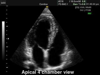
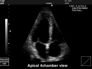
SonoAce-9900. Heart (4 chamber view), B-mode.
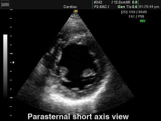
SonoAce-9900. Heart (short axis of LV), B-mode.
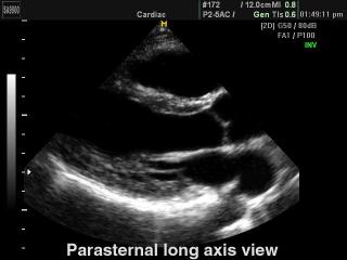
SonoAce-9900. Heart (long axis of LV), B-mode.
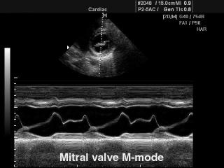
SonoAce-8000. Mitral valve, M-mode.
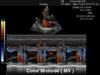
SonoAce-8000. Mitral valve, color M-mode.
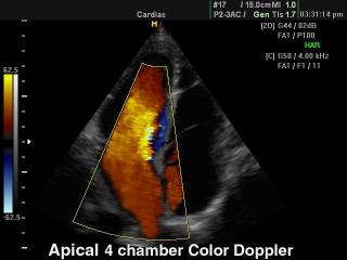
SonoAce-8000. Heart (4 chamber view), color doppler.
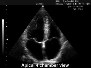
SonoAce-8000. Heart (4 chamber view), B-mode.
SonoAce-8000. Heart (long axis of LV) - inverse harmonic, B-mode.
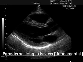
SonoAce-8000. Heart (long axis of LV) - fundamental harmonic, B-mode.
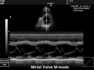
SonoAce-128BW. Mirtal valve, M-mode.
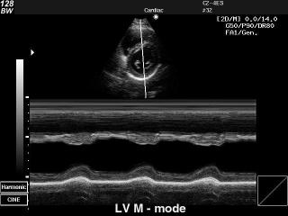
SonoAce-128BW. Heart - left ventricle, M-mode.
SonoAce-128BW. Fetal heart (4 chamber view), B-mode.
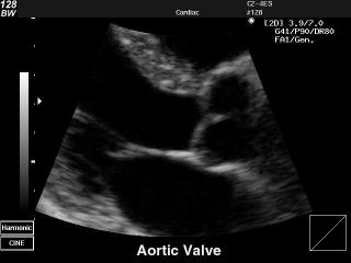
SonoAce-128BW. Aortic valve, B-mode.
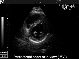
SonoAce-128BW. Heart (short axis of LV), B-mode.
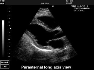
SonoAce-128BW. Heart (long axis of LV), B-mode.
The description of ultrasound examinations in russian (эхография) and in english are situated in the database. The description of the diagnostic in the english language is closer to latin and doctors are likely to prefer this variant.