
Atlas of ultrasound images - abdomen (pg. 6)
In the section "Abdominal ultrasonography" of atlas the results of ultrasound examinations of abdominal cavity organs are represented. Here you can see images of gall bladder, liver, kidney and intestine, the sonograms: ureter stone, intrahepatic calcification, hemangioma, enlarged spleen, etc.
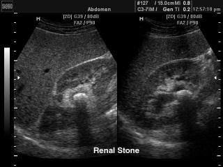
SonoAce-9900. Renal stone, B-mode.
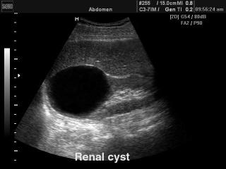
SonoAce-9900. Renal cyst, B-mode.
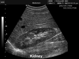
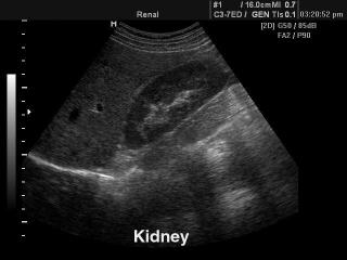
SonoAce-9900. Kidney, B-mode.
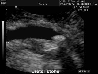
SonoAce-9900. Ureter stone, B-mode.

SonoAce-9900. Spleen - splenomegaly, B-mode.
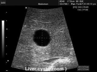
SonoAce-9900. Liver cyst, B-mode.
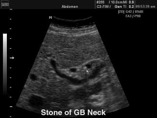
SonoAce-9900. Stone in the neck of gallbladde, B-mode.
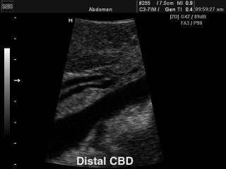
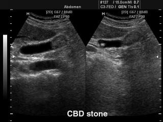
SonoAce-9900. Liver - distal common bile duct, B-mode.
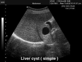
SonoAce-9900. Liver - simple cyst, B-mode.
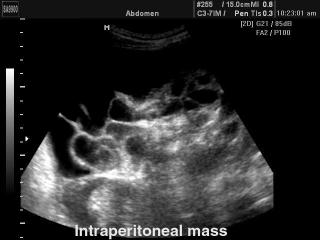
SonoAce-9900. Intestines - intraperitoneal induration, B-mode..
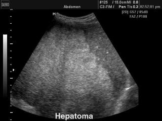
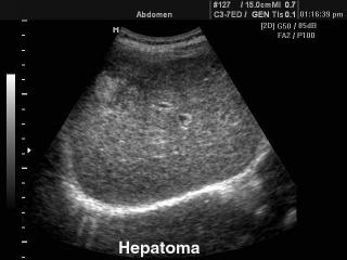
SonoAce-9900. Liver - hepatoma, B-mode.
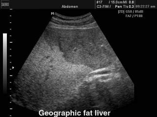
SonoAce-9900. Liver steatosis, B-mode.
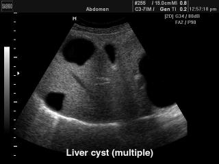
SonoAce-9900. Liver polycystic, B-mode.
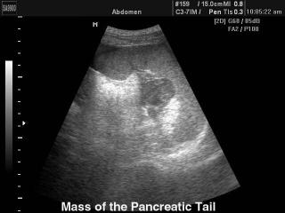
SonoAce-9900. Pancreatic tail mass, B-mode.
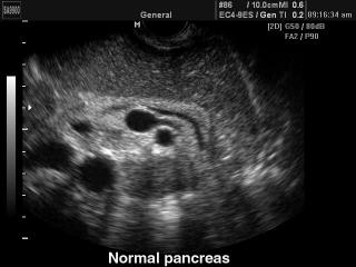
SonoAce-9900. Pancreas - norm, B-mode.
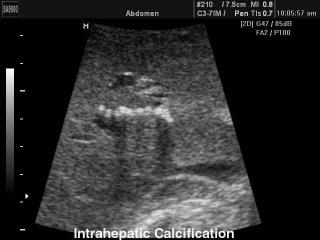
SonoAce-9900. Intrahepatic calcification, B-mode.
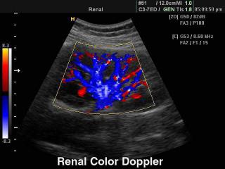
SonoAce-8000. Kidney, color doppler.
SonoAce-8000. Kidney, B-mode.
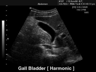
SonoAce-8000. Gall bladder - tissue harmonic, B-mode.
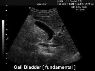
SonoAce-8000. Gall bladder (fundamental harmonic), B-mode.
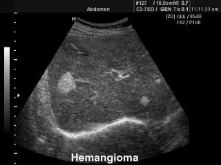
SonoAce-8000. Liver hemangioma, B-mode.
SonoAce-8000. Hepatoma, B-mode.
SonoAce-8000. Gallbladder stone, B-mode.
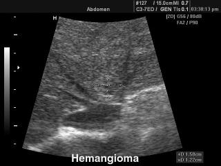
SonoAce-8000. Liver hemangioma, B-mode.
The description of ultrasound examinations in russian (эхография) and in english are situated in the database. The description of the diagnostic in the english language is closer to latin and doctors are likely to prefer this variant.