
Atlas of ultrasound images - abdomen (pg. 2)
In the section "Abdominal ultrasonography" of atlas the results of ultrasound examinations of abdominal cavity organs are represented. Here you can see images of gall bladder, liver, kidney and intestine, the sonograms: ureter stone, intrahepatic calcification, hemangioma, enlarged spleen, etc.
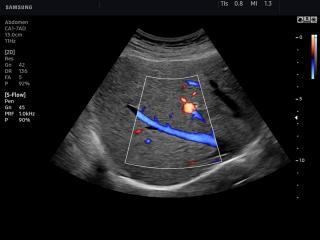
HS50. Liver, S-Flow™.
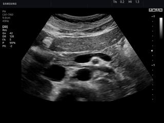
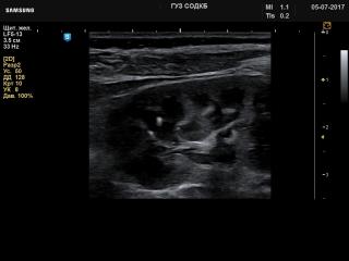
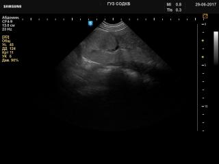
HS50. Pancreas, B-mode.
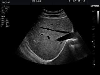
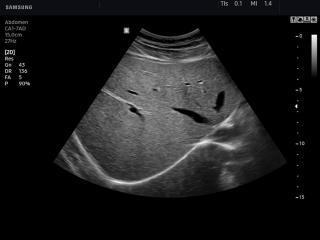
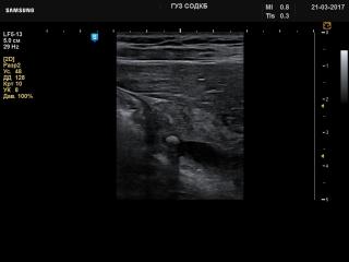
HS50. Liver, B-mode.

HS60. Liver, S-Flow.
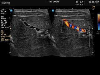
HS60. Pancreas, B-mode.
HS60. Liver, B-mode.
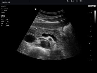
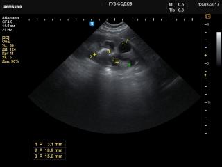
H60. Kidney - small stones, B-mode.
H60. Kidney - stones and dilated renal pelvis, B-mode.
H60. Intestines - intussusception, B-mode.
H60. Kidney - renal dysplasia, B-режим.
H60. Choledochus - gallstone, B-mode.
H60. Liver - autoimmune hepatitis, B-mode & color doppler.
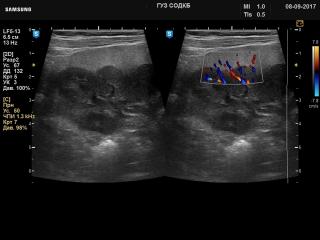
H60. Kidney - nephroblastomatosis, B-mode & color doppler.
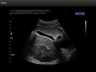
HS70. Liver, S-Harmonic.
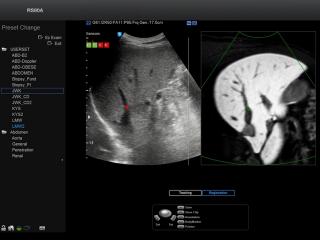
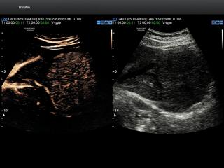
RS80. Liver, S-Fusion.
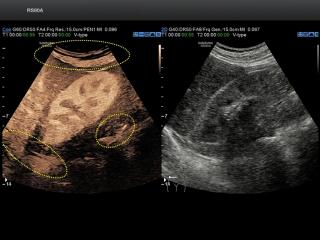
RS80. Liver, S-Fusion with CEUS+.
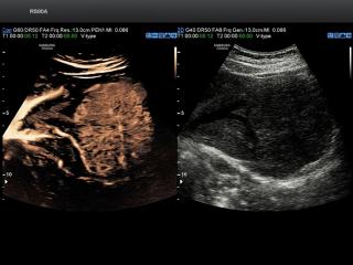
RS80. Liver, CEUS+ and VesselMax.
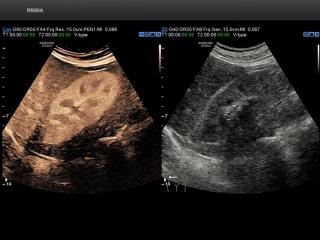
RS80. Liver, CEUS+.
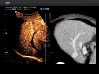
RS80. Kidney, CEUS+ and FlowMax.
RS80. Kidney, CEUS+.
RS80. Liver, S-Harmonic.
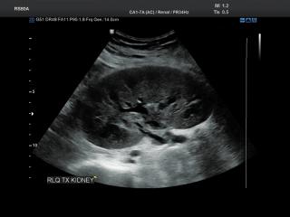
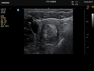
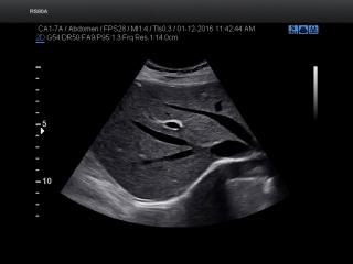
RS80. Renal transplant, B-mode.
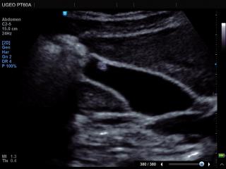
PT60. Liver, ClearVision.
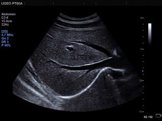
PT60. Gall bladder - polyp, В-mode.
The description of ultrasound examinations in russian (эхография) and in english are situated in the database. The description of the diagnostic in the english language is closer to latin and doctors are likely to prefer this variant.