
Atlas of ultrasound images - abdomen (pg. 3)
In the section "Abdominal ultrasonography" of atlas the results of ultrasound examinations of abdominal cavity organs are represented. Here you can see images of gall bladder, liver, kidney and intestine, the sonograms: ureter stone, intrahepatic calcification, hemangioma, enlarged spleen, etc.
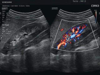
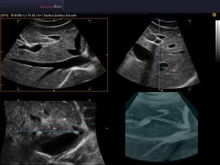
HM70 EVO. Kidney in S-Flow.
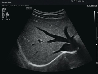
HM70 EVO. Liver, ClearVision.
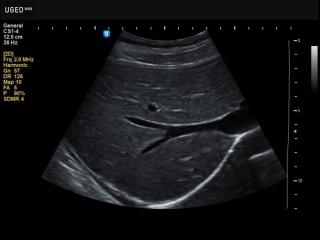
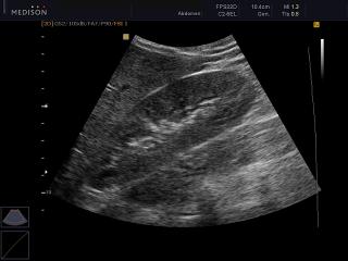
H60. Liver, B-mode.
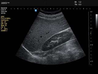
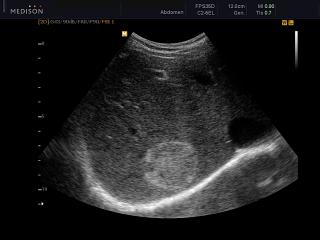
H60. Kidney, B-mode.
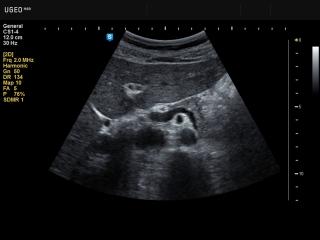
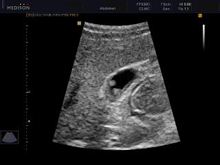
H60. Pancreas, THI.
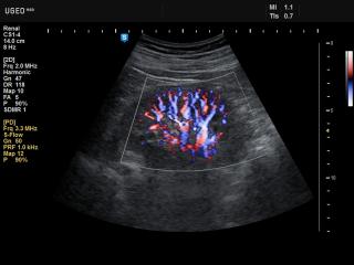
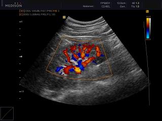
H60. Renal vessels, S-flow.
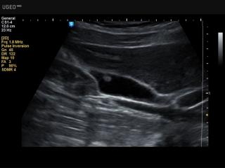
H60. Gall bladder - polyp, SDMR.
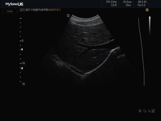
MySono-U6. Gall bladder - polyp, В-mode.
MySono-U6. Liver, B-mode + SRF.
Accuvix-V10. Gallbladde, stone, MSV.
Accuvix-V10. Gallbladder stone, B-mode.
Accuvix-V10. Kidney, color doppler.
Accuvix-V10. Kidney, B-mode.
Accuvix-V10. Liver - hemangioma, B-mode.
Accuvix-A30. Liver, MSV & HDVI.
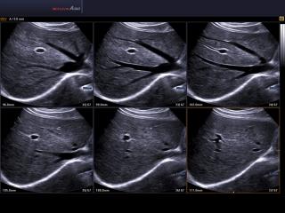
Accuvix-A30. Liver, Multi OVIX, 3D.
Accuvix-A30. Liver, 3D inversion.
Accuvix-A30. Gallbladder stones, Multi OVIX, 3D.
Accuvix-A30. Liver - hemangioma, MSV.
SonoAce-R7. Liver tumor, MSV.
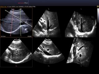
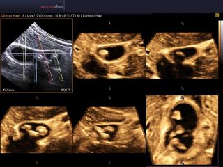
Accuvix-XG. Right kidney, MSV & 3D.
Accuvix-XG. Right kidney, color doppler.
Accuvix-XG. Multiple gallbladder stones, MSV & 3D.
Accuvix-XG. Multiple gallbladder stones, MSV+HDVI & 3D.
The description of ultrasound examinations in russian (эхография) and in english are situated in the database. The description of the diagnostic in the english language is closer to latin and doctors are likely to prefer this variant.