
Atlas of ultrasound images - abdomen (pg. 5)
In the section "Abdominal ultrasonography" of atlas the results of ultrasound examinations of abdominal cavity organs are represented. Here you can see images of gall bladder, liver, kidney and intestine, the sonograms: ureter stone, intrahepatic calcification, hemangioma, enlarged spleen, etc.
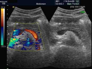
Accuvix-XQ. Blood flow in epigastrio, color doppler (videо).
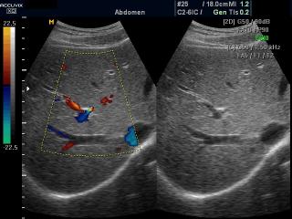
Accuvix-XQ. Liver blood flow, color doppler (videо).
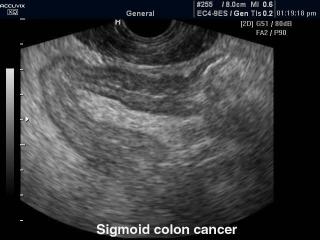
Accuvix-XQ. Sigmoid colon cancer, B-mode.
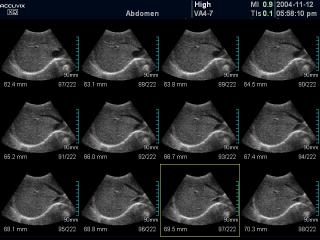
Accuvix-XQ. Kidney, MSV.
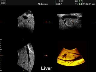
Accuvix-XQ. Liver, MSV.
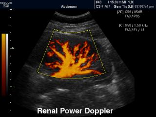
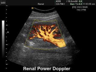
Accuvix-XQ. Kidney, power doppler.
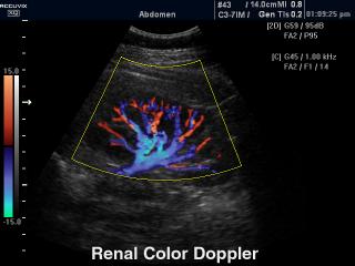
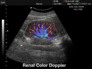
Accuvix-XQ. Kidney, color doppler.
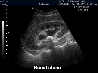
Accuvix-XQ. Renal stone, B-mode.
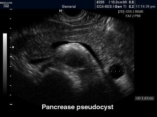
Accuvix-XQ. Pancrease pseudocyst, B-mode.
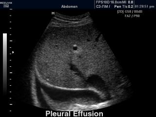
Accuvix-XQ. Pleural effusion, B-mode.
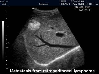
Accuvix-XQ. Liver - metastasis from retroperitoneal lymphoma, B-mode.
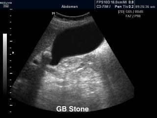
Accuvix-XQ. Gall bladder stone, B-mode.
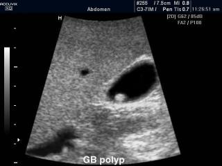
Accuvix-XQ. Gall bladder polyp, B-mode.
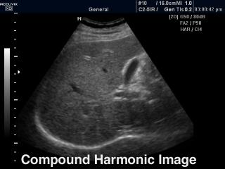
Accuvix-XQ. Liver, compound harmonic mode.
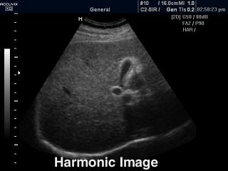
Accuvix-XQ. Liver, tissue harmonic.
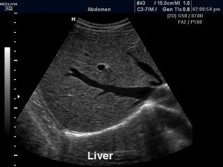
Accuvix-XQ. Liver, B-mode.
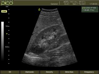
SonoAce-Pico. Kidney, B-mode.
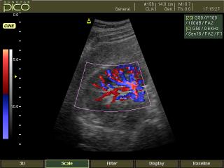
SonoAce-Pico. Kidney, color doppler.
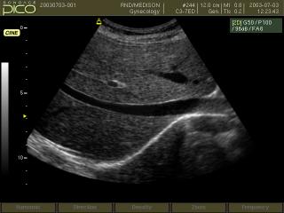
SonoAce-Pico. Liver, B-mode.
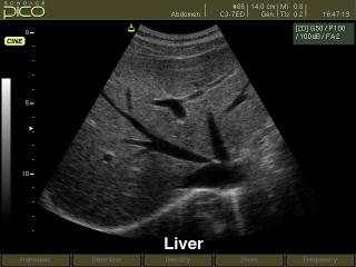
SonoAce-Pico. Liver, B-mode.

SonoAce-6000 CMT. Kidney cancer, B-mode.
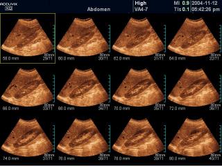
SonoAce-9900. Liver, 3D reconstruction.
SonoAce-9900. Kidney, power doppler.
SonoAce-9900. Kidney, color doppler.
The description of ultrasound examinations in russian (эхография) and in english are situated in the database. The description of the diagnostic in the english language is closer to latin and doctors are likely to prefer this variant.