УЗИ сканер H60 (Samsung Medison, снят с производства)
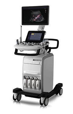
UGEO H60 - универсальный ультразвуковой сканер производства компании Samsung Medison.
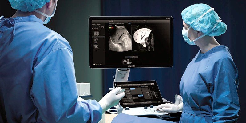
Передовые технологи визуализации и пост-обработки изображений, светодиодный LED монитор с широким углом обзора и сенсорная панель управления. Компактный дизайн позволяет разместить UGEO H60 возле постели больного для максимального удобства. Рекомендуется применение сканера в современных диагностических центрах и медицинских исследовательских институтах.
Базовая комплектация: сканер UGEO H60 (встроенные модули: цветного, энергетического, направленного энергетического и импульсно-волнового допплера, тканевая гармоника, SonoView; кинопамять; USB-порты, встроенная клавиатура с подсветкой и трекболом, четыре активных порта, гибридный бимформер (блок формирования изображения), флакон геля 250 мл и руководство оператора).
Опции для сканера UGEO H60: система Live 3D; кардиопакет; ЭКГ модуль; панорамное сканирование; постоянно-волновой допплер; опции 3D XI, SDMR, CEUS+, AutoIMT, SCI; система DICOM, педаль дистанционного управления; нагреватель геля.
Видео-презентация сканера UGEO H60
Области применения: акушерство и гинекология, абдоминальные исследования и маммология, урология и эхокардиография, поверхностно расположенные органы и исследования сосудов, мускуло-скелетные исследования, а также нефрология, онкология и транскраниальная допплерография.
Основные характеристики УЗ сканера UGEO H60
- Стационарный ультразвуковой сканер.
- Широкоформатный LED монитор (монитор с диодной подсветкой) высокого разрешения, с широким углом обзора.
- Сенсорная панель управления (touch-screen).
- Кинопамять - автоматическая видео-запись фрагмента исследования с возможностями "перемотки", редактирования, проведения расчетов и последующей записи видео в файл.
- Разъемы для одновременного подключения до 5-х датчиков (4 + 1 CW).
- USB-порты (для подключения периферических устройств, внешних накопителей: флеш-карт).
- Система SonoView - система архивации и дальнейшего просмотра статических и динамических изображений (база данных изображений), имеется возможность копирования изображений на внешние накопители (подключение по USB), проводить измерения в архиве.
Режимы визуализации
- B - двухмерное сканирование в оттенках серой шкалы, тканевая гармоника (в том числе пульс-инверсная).
- M - одномерный режим для исследования сердца, анатомический М-режим (необходим кардиопакет), цветной М-режим (необходим кардиопакет).
- CD - цветное допплеровское картирование.
- PD - энергетический допплер.
- S-Flow - инновационная технология цветового картирования кровотока позволяет увидеть и детально оценить кровоток в мельчайших периферических сосудах, что гарантирует наиболее точную диагностику в самых сложных случаях.
- TDI - тканевый допплер (необходим кардиопакет).
- PW - импульсно-волновой допплер.
- Steering - изменение допплеровского угла в режимах CD и PD, изменение угла сканирования в В-режиме на линейных датчиках.
- HPRF - высокочастотный импульсно-волновой допплер.
- CW - постоянно-волновой допплер (опция).
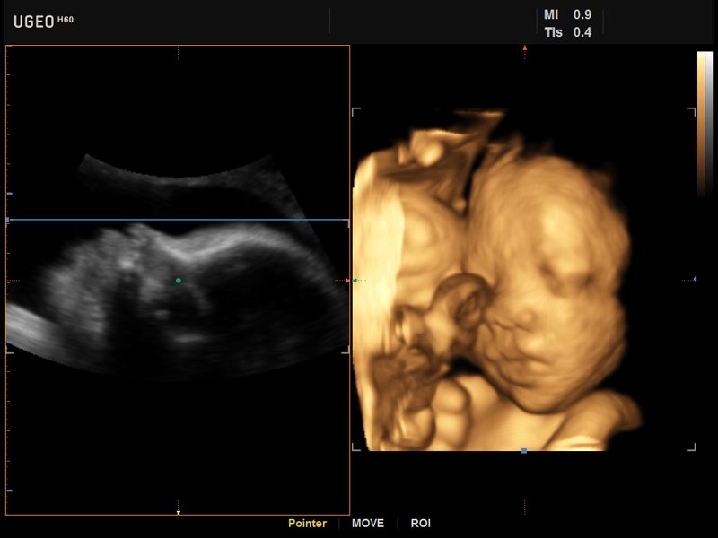
- 4D - трехмерное сканирование объемными датчиками в реальном масштабе времени (необходима опция "Live 3D").
- Режимы одновременного отображения на экране 2-х, 4-х и более изображений, в т.ч. изображений в режимах B/C, B/PD в реальном масштабе времени.
- Смешанные режимы (B/M, B/PWD, B/C, B/PD, B/PD/PWD, B/C/PWD).
- Трапециевидный режим (для линейных датчиков).
- Масштабирование.
Опции
- Система Live 3D - трехмерное сканирование объемными датчиками в реальном масштабе времени (4D).
- Кардиопакет: тканевый допплер (TDI) + анатомический М-режим + цветной М-режим (CM) + программы расчетов.
- Модуль ЭКГ.
- Модуль панорамного сканирования.
- Модуль CW - постоянно-волновой допплер.
- Пакет опций 3D XI (объемная ультразвуковая томография).
- Модуль VOCAL - автоматический расчет объемов, объемных гистограмм и сосудистых индексов (VI, FI, VFI) объемных изображений.
- Модуль SDMR (Samsung Dynamic Magnetic Resonance) - программа оптимизации изображения по магнитно-резонансному алгоритму для усиления контрастного разрешения и улучшения четкости контуров.
- Модуль VolumeNT&IT - полуавтоматическое измерение толщины воротникового пространства (маркер синдрома Дауна) и интракраниального пространства (четвертый желудочек, маркер спина бифида) в объемном изображении.
- Модуль CEUS+ (Contrast Enchansment UltraSound) - исследования с применением контрастных веществ.
- Модуль AutoIMT - автоматическое вычисление комплекса интима-медиа общей сонной артерии (Intima Media Thickness). Данная оценка имеет большое значение для ранней диагностики атеросклероза и оценки риска развития инсульта и инфаркта миокарда.
- Модуль SCI (Spatial Compound Imaging) - детализация изображения и уменьшение артефактов за счет технологии получения изображения с учетом нескольких углов инсонации.
- Система DICOM - возможность сетевой интеграции с PACS-системами (например, для архивации или печати ультразвуковых эхограмм на оборудовании других производителей медтехники).
- Педаль дистанционного управления.
- Нагреватель геля.
Пакет опций 3D XI (объемная ультразвуковая томография)
- MSV (Multi-Slice View или мультислайсинг) - возможность одновременного просмотра на экране множественных срезов, полученных при трехмерном сканировании.
- VolumeCT - трехмерная реконструкция изображений в виде куба (Cube Sectional View) или трех пересекающихся плоскостей (Cross View).
- OVIX (Oblique View eXtended) - получение фрагмента трехмерного изображения (в виде нескольких полупрозрачных сканов, последовательно наложенных один на другой) в направлении произвольного косого среза трехмерного объекта исследования.
Инновационные технологии
- Quick Scan - режим автоматической настройки изображения (нажатием одной кнопки) исследуемого органа в B- и D-режиме (настройка оптимальных параметров и фильтров за счет автоматического распознавания исследуемого органа по интеллектуальной базе данных человеческих органов).
- SRF (Speckle Reduction Filter) - фильтр подавления шума.
- VCE (Volume Contrast Enhancement) - функция усиления качества изображения трехмерного объекта за счёт удаления зон с неотчётливой визуализацией.
- VSI (Volume Shade Imaging) - "визуализация объемных оттенков" на 3D-изображении.
- FSI (Full Spectrum Imaging) - многоступенчатый алгоритм получения избирательного контрастирования изображения по всей глубине сканирования.
- Digital TGC - цифровая регулировка усиления по глубине.
- See-Thru - технология, использующая объединение трехмерного энергетического допплера и серошкального 3D изображения.
- Технология доступа к необработанным ("сырым") данным, сохраненным в архиве с возможностью изменения параметров сканирования.
Основные измерения
- В-режим: расстояние, периметр, угол, площадь, эллипс, окружность, объем.
- D-режим: скорость, давление, ускорение, замедление.
- M-режиме: время, расстояние, уклон.
Пакеты расчетов (измерения и отчеты)
- Гинекология: матка, левый и правый яичники, левый и правый фолликулы, левая и правая яичниковые артерии, левая и правая маточные артерии, эндометрий, киста, опухоль, объемное образование и др.
- Акушерство: биометрия плода (плодное яйцо (GS), теменно-копчиковая длина (CRL), бипариетальный размер головки (BPD), лобно-затылочное расстояние (OFD), окружности головы (НC), передне-задний размер живота (APD), поперечный размер живота (TAD), окружность живота (AC), длина бедра (FL) и др.), длинные кости плода (плечевая (Humerus), локтевая (Ulna), лучевая (Rad), большеберцовая (Tibia), малая берцовая, ключица (Clav) и позвоночник (LV), краниологическое исследование плода (мозжечок (CEREB), внешнее (OOD) и внутреннее (IOD) межглазничные расстояния, большая цистерна, шейная складка, боковые желудочки, носовая кость), другие показатели плода (ступня, ухо, средняя фаланга, почки, таз), индекс околоплодных вод (AFI), допплерометрия (пупочная артерия, средняя мозговая артерия, маточные артерии, плацентарная артерия, сонные артерии, аорта плода, венозный проток, ЧСС плода); уравнения для оценки веса плода (Хедлок (Hadlock) 1-4, Хансман (Hansmann) и Мерц (Merz)); таблицы, определяемые пользователем.
- Сердце плода: измерения в В-режиме (отношение площади сердца и грудной клетки), измерения в М-режиме (толщина межжелудочковой перегородки в диастолу, конечнодиастолический размер левого желудочка, толщина задней стенки левого желудочка в диастолу, толщина межжелудочковой перегородки в систолу, размер левого желудочка в систолу, толщина задней стенки левого желудочка в систолу, внутренний размер правого желудочка в диастолу), измерения в режиме спектрального допплера (легочный ствол, артериальный проток, нижняя полая вена, венозный проток, восходящая аорта, нисходящая аорта, трансмитральный кровоток, митральная регургитация, трикуспидальный кровоток, трикуспидальная регургитация, индекс преднагрузки, ЧСС).
- Пакет кардиологических исследований.
М-режим: измерение диаметра аорты, передне-заднего размера ЛП, толщины МЖП (систолическая и диастолическая), толщины ЗСЛЖ (систолическая и диастолическая), размеров ЛЖ и ПЖ (систолический и диастолический), ФВ (Teichholz).
B-режим: измерение диаметра аорты (восходящей, дуги, нисходящей, на уровне синусов Вальсальвы, на уровне створок аортального клапана), определение размеров ЛП и ПП (максимальный, минимальный, систолический, диастолический, переднее-задний, верхнее-нижний, медиально-латеральный), расчет объемов ЛП и ПП, объемов ЛЖ (метод "Площадь-Длина", метод дисков (Simpson)), массы миокарда ЛЖ, индекса массы миокарда ЛЖ.
CD-режим (ЦДК): измерение радиуса ПФСМР (PISA), полуколичественная оценка трансмитрального, транстрикуспидального, трансаортального и транспульмонального кровотока (оценка регургитации), оценка аномальных сбросов крови через МПП И МЖП.
PW-режим (импульсно-волновой допплер): автоматическая, полуавтоматическая и ручная трассировка допплеровского спектра митрального, аортального и трикуспидального клапанов, клапана легочной артерии, кровотока в выходном тракте ЛЖ и ПЖ (пиковая/средняя скорость, пиковый/средний градиент давления, время изоволюметрического расслабления ЛЖ, время ускорения, замедления, выброса), оценка кровотока легочных и печеночных вен.
CW-режим (постоянно-волновой допплер): программы расчета работы митрального, аортального и трикуспидального клапанов, клапана легочной артерии.
TD-режим (тканевой допплер): количественная оценка локальной сократительной функции стенок ЛЖ и ПЖ. - Сонные артерии: автоматическая, полуавтоматическая, ручная трассировка доплеровского спектра; ПСС, КДС, %СтПлощ, %Ст Диам, площадь сосуда, диаметр сосуда, средняя толщина интимы, объемный кровоток.
- Артерии верхних и нижних конечностей: автоматическая, полуавтоматическая, ручная трассировка доплеровского спектра; ПСС, КДС, %СтПлощ, %Ст Диам, площадь сосуда, диаметр сосуда, объемный кровоток.
- Вены верхних и нижних конечностей: автоматическая, полуавтоматическая, ручная трассировка доплеровского спектра; максимальная скорость, диаметр сосуда.
- Сосуды брюшной полости: автоматическая, полуавтоматическая, ручная трассировка доплеровского спектра; ПСС, КДС, %СтПлощ, %Ст Диам, площадь сосуда, диаметр сосуда, объемный кровоток.
- Урология: размеры почек, объем мочевого пузыря, остаточный объем, объем предстательной железы по WG, объем Т-зон, объем почки (методы измерения объема: три расстояния, три расстояния и коэффициент, эллипсоид).
- Измерение угла тазобедренного сустава (новорожденные).
- Щитовидная железа.
- Молочная железа.
Сокращения: ЛП/ПП - левое/правое предсердие, МЖП - межжелудочковая перегородка, МПП - межпредсердная перегородка, ЗСЛЖ - задняя стенка левого желудочка, ЛЖ/ПЖ - левый/правый желудочек, ФВ - фракция выброса, ПФСМР - площадь формирующейся струи митральной регуритации (PISA - proximal isovelocity surface area), ПСС/КДС - пиковая систолическая / конечная диастолическая скорость.
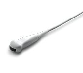
Датчики для сканера UGEO H60
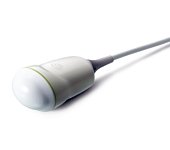
Конвексные датчики
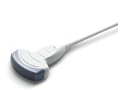
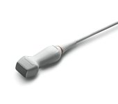
Конвексный датчик 1-4 МГц
Акушерские исследования (плод, сердце плода), гинекология (матка, яичники), абдоминальные исследования (печень, желчный пузырь, поджелудочная железа, селезенка, глубокие сосуды), почки.
Биопсийный набор: есть.
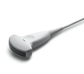
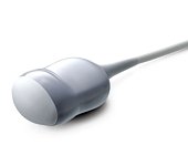
Конвексный датчик 2-8 МГц
Акушерские исследования (плод, сердце плода), гинекология (матка, яичники), абдоминальные исследования (печень, желчный пузырь, поджелудочная железа, селезенка, глубокие сосуды), почки.
Биопсийный набор: есть.
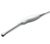
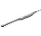
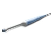
Конвексный датчик 4-9 МГц (вагинальный)
Акушерские исследования (ранние сроки), гинекология (матка, яичники), урология (предстательная железа), исследования прямой кишки.
E-motion Marker: есть
Биопсийный набор: есть.
Конвексный датчик 4-9 МГц (неонатальный)
Неонатология и педиатрия: абдоминальные исследования, почки, сердце, глубокие сосуды, мозг.
Биопсийный набор: нет.
Конвексный датчик 4-9 МГц (ректо-вагинальный)
Акушерские исследования (ранние сроки), гинекология (матка, яичники), урология (предстательная железа), исследования прямой кишки.
Биопсийный набор: есть.
Фазированные датчики
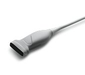
Фазированный датчик 2-4 МГц
Кардиология и транскраниальные исследования у взрослых.
Биопсийный набор: нет.
Фазированный датчик 3-8 МГц
Детская кардиология, транскраниальные исследования в педиатрии.
Биопсийный набор: нет.
Линейные датчики
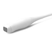
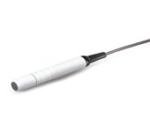
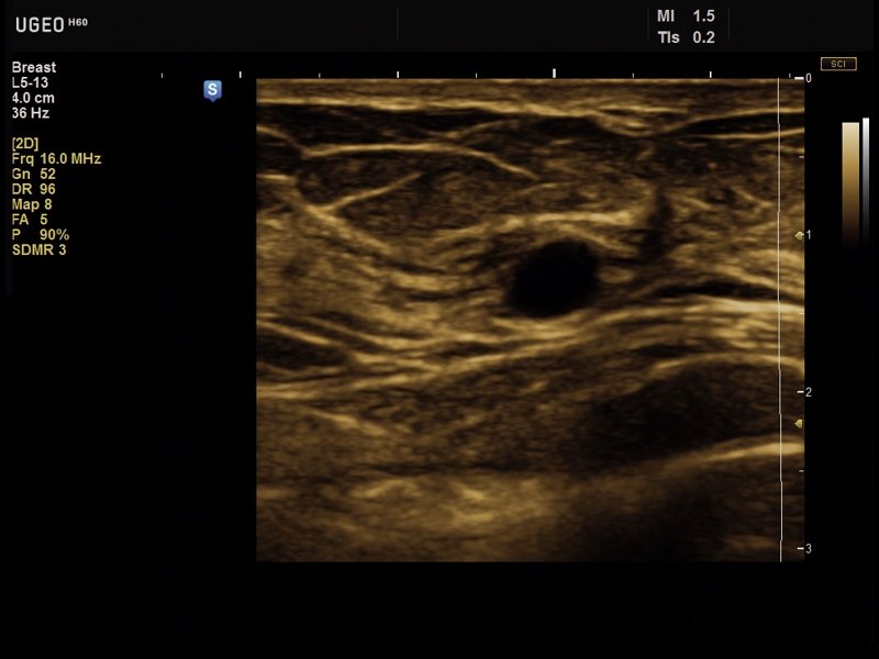
Линейный датчик 5-13 МГц
Поверхностные структуры (щитовидная железа, молочная железа, лимфоузлы), мускулоскелетные исследования (суставы, мышцы, подкожные структуры), периферические сосуды.
Биопсийный набор: есть.
Объемные датчики
Объемный датчик 2-6 МГц
Трехмерные абдоминальные исследования, акушерство и гинекология.
Биопсийный набор: есть.
Объемный датчик 4-8 МГц
Трехмерные абдоминальные исследования, акушерство (трехмерное УЗИ плода) и гинекология.
Биопсийный набор: есть.
Объемный датчик 4-9 МГц (ректо-вагинальный)
Трехмерные исследования в акушерстве (ранние сроки), гинекологии (матка, яичники), урологии (предстательная железа), исследования прямой кишки.
Биопсийный набор: есть.
Допплеровские датчики

Допплеровский датчик 2.0 МГц (слепой допплер)
Транскраниальные исследования, сосуды.
Биопсийный набор: нет.
Допплеровский датчик 4.0 МГц (слепой допплер)
Транскраниальные исследования, сосуды.
Биопсийный набор: нет.
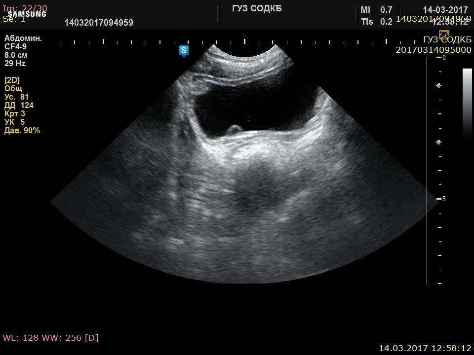
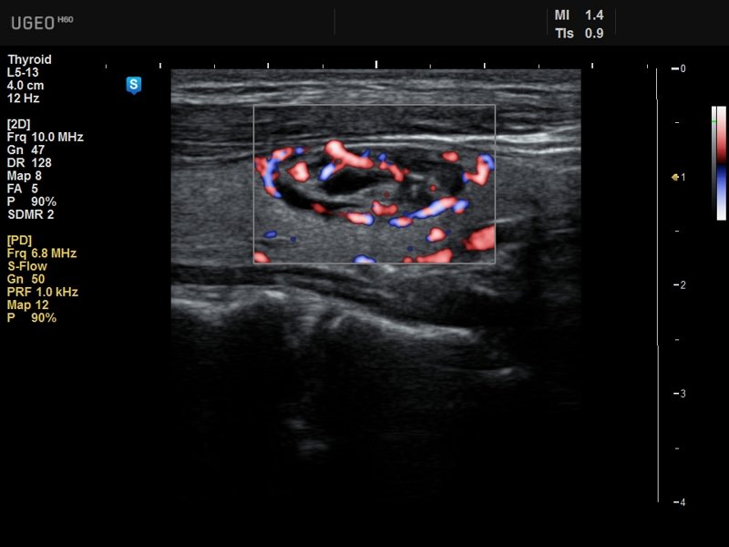
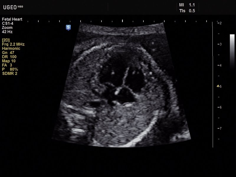
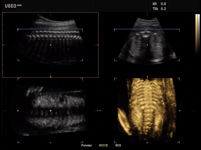
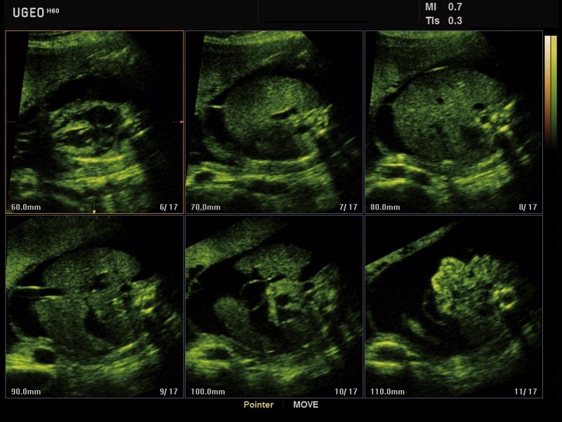
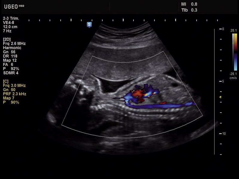
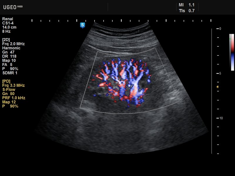
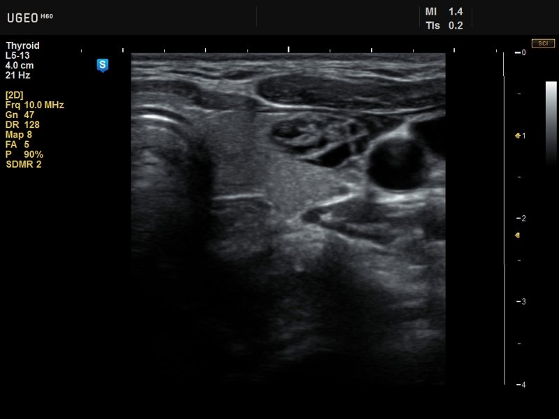
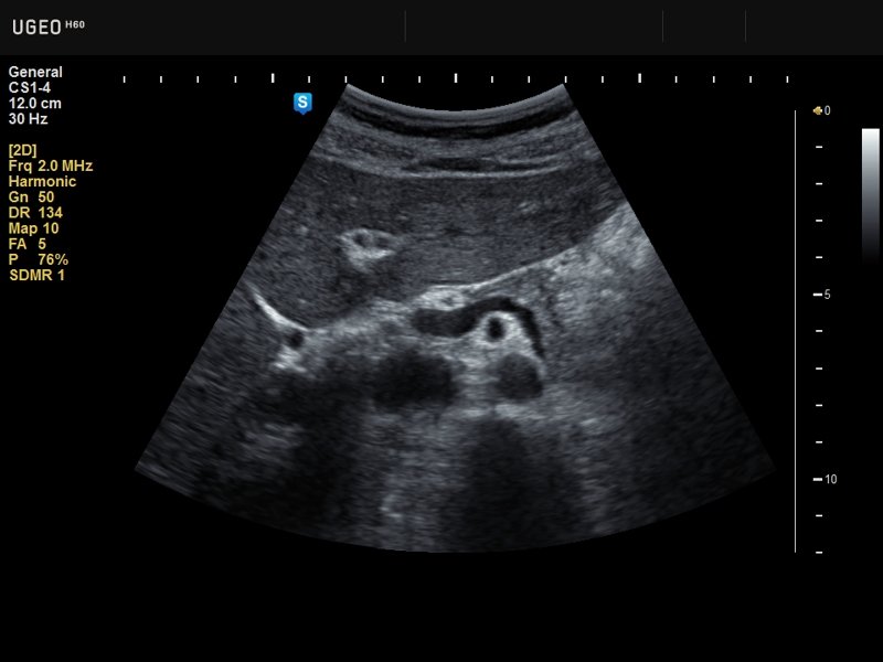
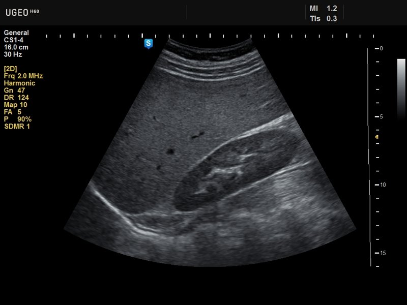
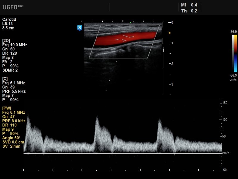
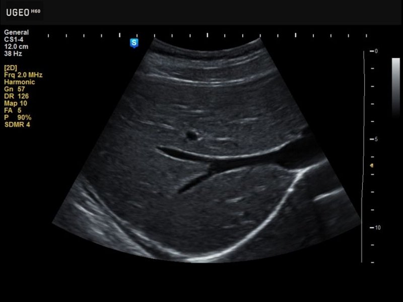
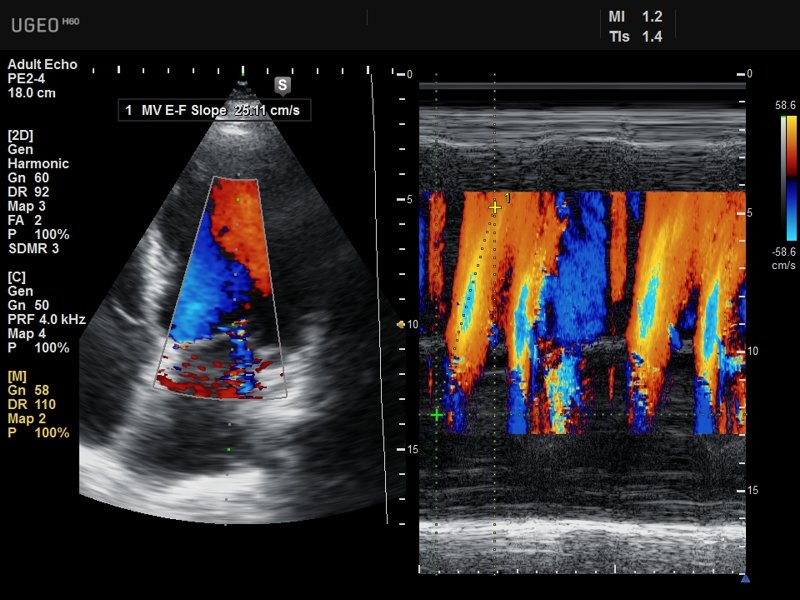
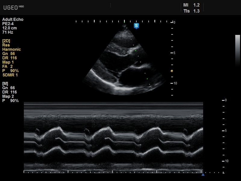
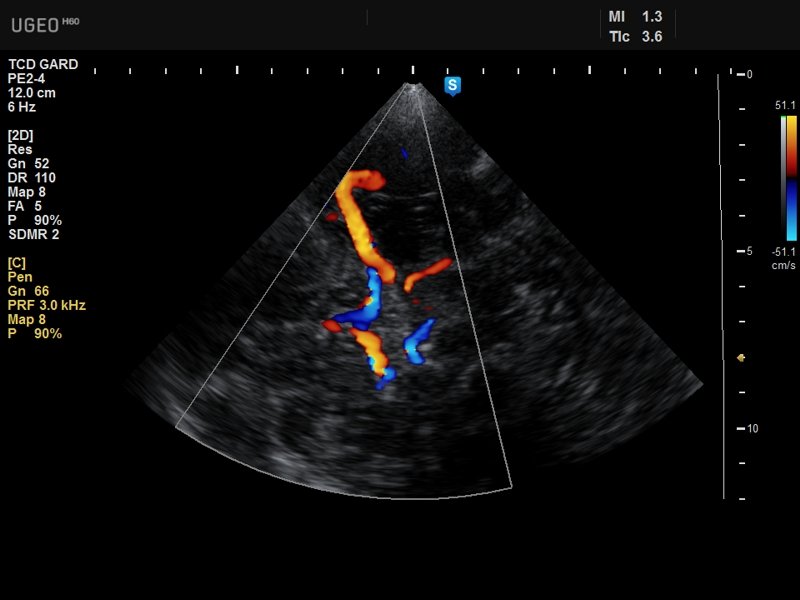
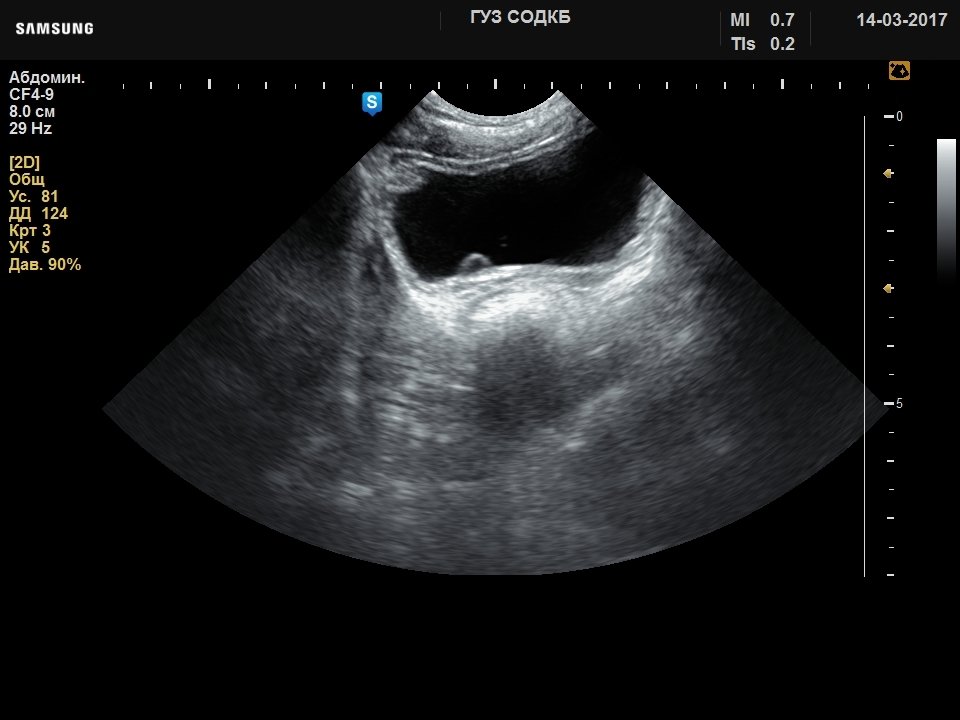
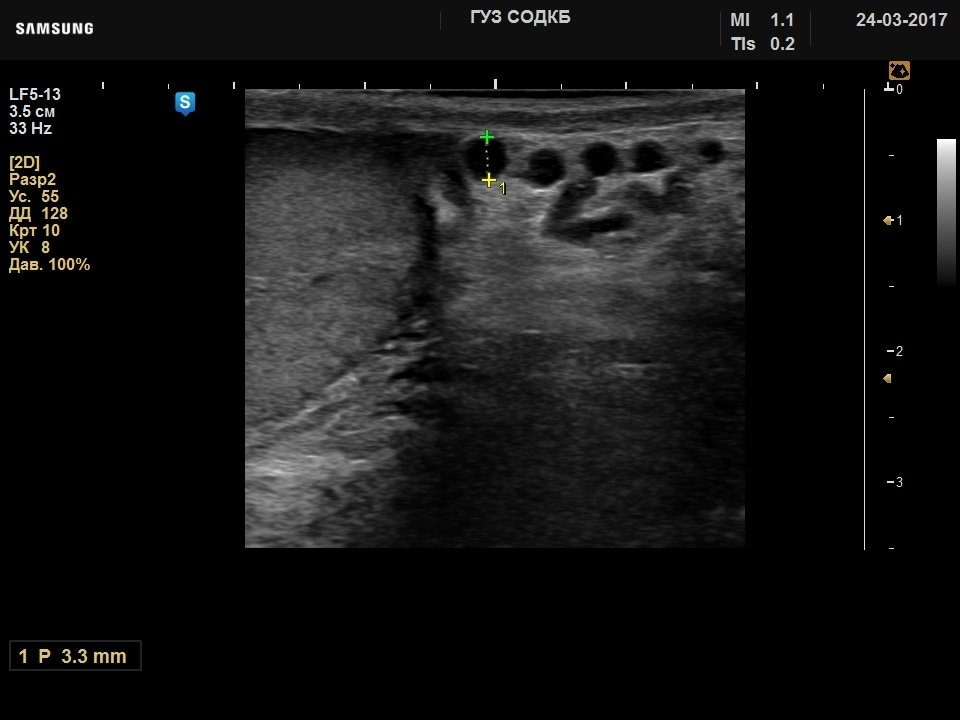
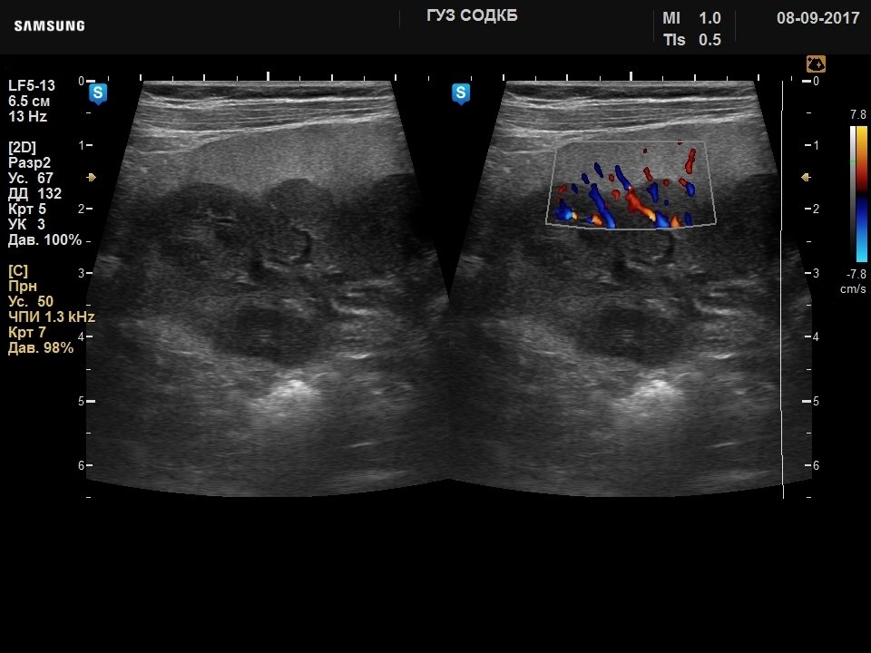
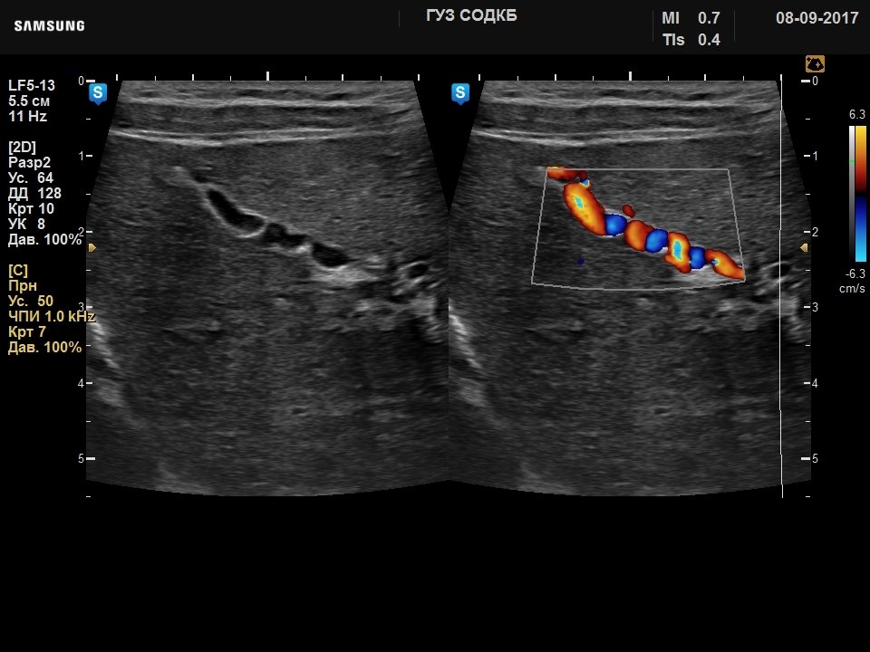
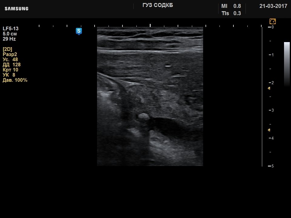
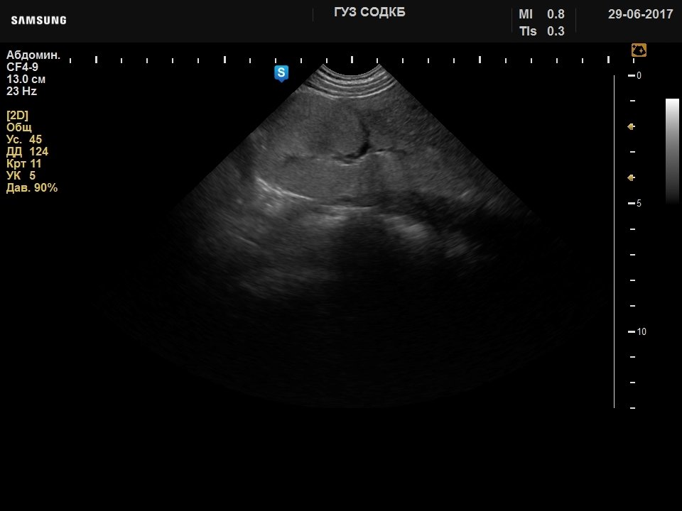
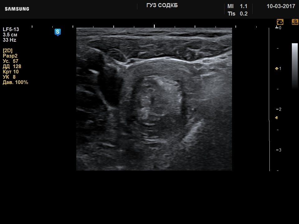
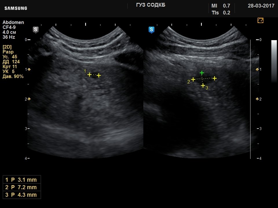
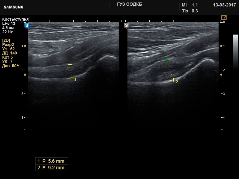
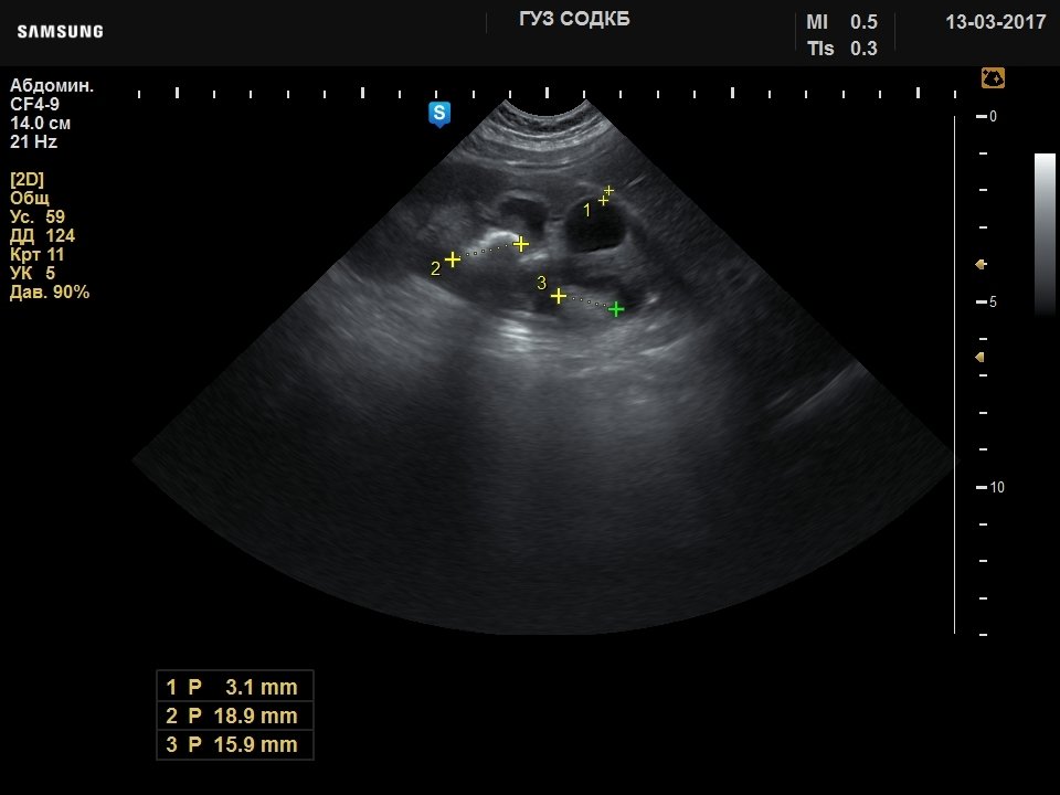
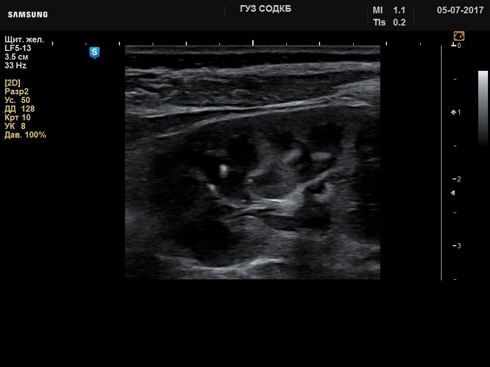
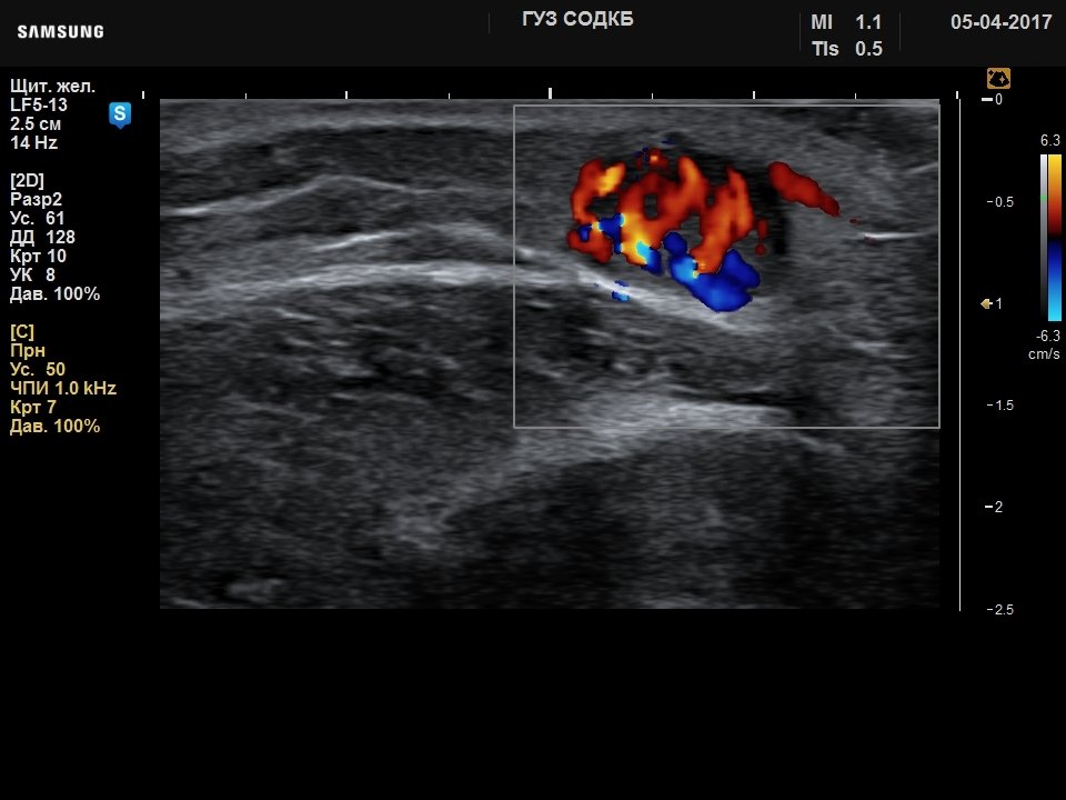
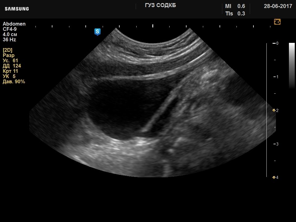
Примеры УЗИ на сканере UGEO H60 (эхограммы)


Внимание! Копирование или частичная перепечатка материалов проспекта сканера "UGEO H60" на русском языке и размещение в интернет без согласия владельца (ЗАО "Медиэйс") запрещена, запрос на копирование материала можно отправить здесь.